Calcium »
PDB 3pe0-3pty »
3pr4 »
Calcium in PDB 3pr4: DPO4 Y12A Mutant Incorporating Dadp Opposite Template Dt
Enzymatic activity of DPO4 Y12A Mutant Incorporating Dadp Opposite Template Dt
All present enzymatic activity of DPO4 Y12A Mutant Incorporating Dadp Opposite Template Dt:
2.7.7.7;
2.7.7.7;
Protein crystallography data
The structure of DPO4 Y12A Mutant Incorporating Dadp Opposite Template Dt, PDB code: 3pr4
was solved by
K.N.Kirouac,
Z.Suo,
H.Ling,
with X-Ray Crystallography technique. A brief refinement statistics is given in the table below:
| Resolution Low / High (Å) | 26.11 / 2.65 |
| Space group | P 21 21 2 |
| Cell size a, b, c (Å), α, β, γ (°) | 96.347, 102.004, 52.226, 90.00, 90.00, 90.00 |
| R / Rfree (%) | 19.7 / 25.5 |
Calcium Binding Sites:
The binding sites of Calcium atom in the DPO4 Y12A Mutant Incorporating Dadp Opposite Template Dt
(pdb code 3pr4). This binding sites where shown within
5.0 Angstroms radius around Calcium atom.
In total 3 binding sites of Calcium where determined in the DPO4 Y12A Mutant Incorporating Dadp Opposite Template Dt, PDB code: 3pr4:
Jump to Calcium binding site number: 1; 2; 3;
In total 3 binding sites of Calcium where determined in the DPO4 Y12A Mutant Incorporating Dadp Opposite Template Dt, PDB code: 3pr4:
Jump to Calcium binding site number: 1; 2; 3;
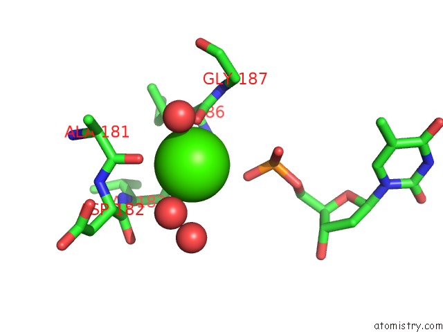
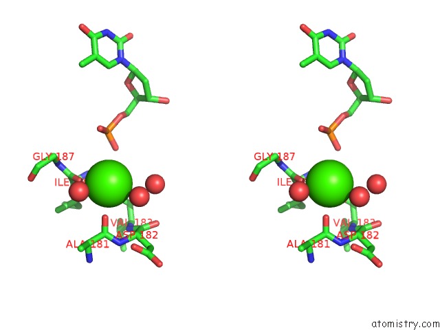
Calcium binding site 1 out of 3 in 3pr4
Go back to
Calcium binding site 1 out
of 3 in the DPO4 Y12A Mutant Incorporating Dadp Opposite Template Dt
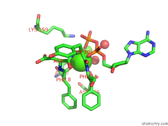
Mono view
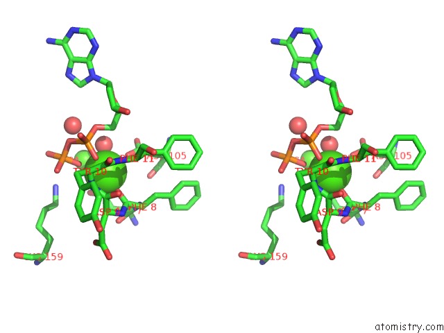
Stereo pair view
Mono view
Stereo pair view
A full contact list of Calcium with other atoms in the Ca binding
site number 1 of DPO4 Y12A Mutant Incorporating Dadp Opposite Template Dt within 5.0Å range:
|
Calcium binding site 2 out of 3 in 3pr4
Go back to
Calcium binding site 2 out
of 3 in the DPO4 Y12A Mutant Incorporating Dadp Opposite Template Dt
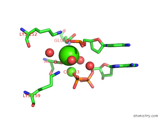
Mono view
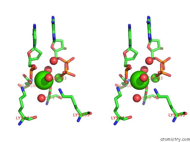
Stereo pair view
Mono view
Stereo pair view
A full contact list of Calcium with other atoms in the Ca binding
site number 2 of DPO4 Y12A Mutant Incorporating Dadp Opposite Template Dt within 5.0Å range:
|
Calcium binding site 3 out of 3 in 3pr4
Go back to
Calcium binding site 3 out
of 3 in the DPO4 Y12A Mutant Incorporating Dadp Opposite Template Dt
Mono view
Stereo pair view
Mono view
Stereo pair view
A full contact list of Calcium with other atoms in the Ca binding
site number 3 of DPO4 Y12A Mutant Incorporating Dadp Opposite Template Dt within 5.0Å range:
|
Reference:
K.N.Kirouac,
Z.Suo,
H.Ling.
Structural Mechanism of Ribonucleotide Discrimination By A Y-Family Dna Polymerase. J.Mol.Biol. V. 407 382 2011.
ISSN: ISSN 0022-2836
PubMed: 21295588
DOI: 10.1016/J.JMB.2011.01.037
Page generated: Sat Jul 13 16:55:07 2024
ISSN: ISSN 0022-2836
PubMed: 21295588
DOI: 10.1016/J.JMB.2011.01.037
Last articles
Zn in 9J0NZn in 9J0O
Zn in 9J0P
Zn in 9FJX
Zn in 9EKB
Zn in 9C0F
Zn in 9CAH
Zn in 9CH0
Zn in 9CH3
Zn in 9CH1