Calcium »
PDB 3pe0-3pty »
3psr »
Calcium in PDB 3psr: Human Psoriasin (S100A7) CA2+ Bound Form (Crystal Form I)
Protein crystallography data
The structure of Human Psoriasin (S100A7) CA2+ Bound Form (Crystal Form I), PDB code: 3psr
was solved by
D.E.Brodersen,
J.Nyborg,
M.Kjeldgaard,
with X-Ray Crystallography technique. A brief refinement statistics is given in the table below:
| Resolution Low / High (Å) | 100.00 / 2.50 |
| Space group | P 21 21 21 |
| Cell size a, b, c (Å), α, β, γ (°) | 52.150, 56.670, 76.380, 90.00, 90.00, 90.00 |
| R / Rfree (%) | 22 / 29.5 |
Other elements in 3psr:
The structure of Human Psoriasin (S100A7) CA2+ Bound Form (Crystal Form I) also contains other interesting chemical elements:
| Zinc | (Zn) | 1 atom |
Calcium Binding Sites:
The binding sites of Calcium atom in the Human Psoriasin (S100A7) CA2+ Bound Form (Crystal Form I)
(pdb code 3psr). This binding sites where shown within
5.0 Angstroms radius around Calcium atom.
In total 2 binding sites of Calcium where determined in the Human Psoriasin (S100A7) CA2+ Bound Form (Crystal Form I), PDB code: 3psr:
Jump to Calcium binding site number: 1; 2;
In total 2 binding sites of Calcium where determined in the Human Psoriasin (S100A7) CA2+ Bound Form (Crystal Form I), PDB code: 3psr:
Jump to Calcium binding site number: 1; 2;
Calcium binding site 1 out of 2 in 3psr
Go back to
Calcium binding site 1 out
of 2 in the Human Psoriasin (S100A7) CA2+ Bound Form (Crystal Form I)
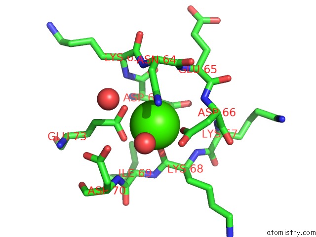
Mono view
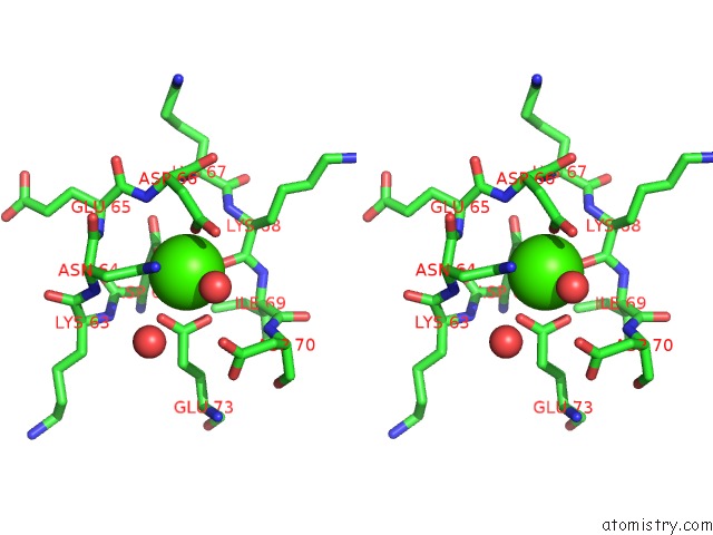
Stereo pair view
Mono view
Stereo pair view
A full contact list of Calcium with other atoms in the Ca binding
site number 1 of Human Psoriasin (S100A7) CA2+ Bound Form (Crystal Form I) within 5.0Å range:
|
Calcium binding site 2 out of 2 in 3psr
Go back to
Calcium binding site 2 out
of 2 in the Human Psoriasin (S100A7) CA2+ Bound Form (Crystal Form I)
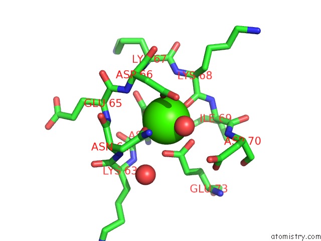
Mono view
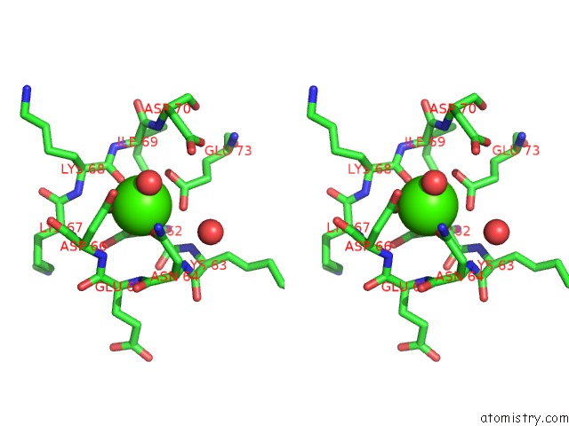
Stereo pair view
Mono view
Stereo pair view
A full contact list of Calcium with other atoms in the Ca binding
site number 2 of Human Psoriasin (S100A7) CA2+ Bound Form (Crystal Form I) within 5.0Å range:
|
Reference:
D.E.Brodersen,
J.Nyborg,
M.Kjeldgaard.
Zinc-Binding Site of An S100 Protein Revealed. Two Crystal Structures of CA2+-Bound Human Psoriasin (S100A7) in the ZN2+-Loaded and ZN2+-Free States. Biochemistry V. 38 1695 1999.
ISSN: ISSN 0006-2960
PubMed: 10026247
DOI: 10.1021/BI982483D
Page generated: Tue Jul 8 15:34:10 2025
ISSN: ISSN 0006-2960
PubMed: 10026247
DOI: 10.1021/BI982483D
Last articles
Fe in 2YXOFe in 2YRS
Fe in 2YXC
Fe in 2YNM
Fe in 2YVJ
Fe in 2YP1
Fe in 2YU2
Fe in 2YU1
Fe in 2YQB
Fe in 2YOO