Calcium »
PDB 3pe0-3pty »
3ptn »
Calcium in PDB 3ptn: On the Disordered Activation Domain in Trypsinogen. Chemical Labelling and Low-Temperature Crystallography
Enzymatic activity of On the Disordered Activation Domain in Trypsinogen. Chemical Labelling and Low-Temperature Crystallography
All present enzymatic activity of On the Disordered Activation Domain in Trypsinogen. Chemical Labelling and Low-Temperature Crystallography:
3.4.21.4;
3.4.21.4;
Protein crystallography data
The structure of On the Disordered Activation Domain in Trypsinogen. Chemical Labelling and Low-Temperature Crystallography, PDB code: 3ptn
was solved by
J.Walter,
W.Steigemann,
T.P.Singh,
H.Bartunik,
W.Bode,
R.Huber,
with X-Ray Crystallography technique. A brief refinement statistics is given in the table below:
| Resolution Low / High (Å) | 6.50 / 1.70 |
| Space group | P 31 2 1 |
| Cell size a, b, c (Å), α, β, γ (°) | 55.110, 55.110, 109.380, 90.00, 90.00, 120.00 |
| R / Rfree (%) | 19.8 / n/a |
Calcium Binding Sites:
The binding sites of Calcium atom in the On the Disordered Activation Domain in Trypsinogen. Chemical Labelling and Low-Temperature Crystallography
(pdb code 3ptn). This binding sites where shown within
5.0 Angstroms radius around Calcium atom.
In total only one binding site of Calcium was determined in the On the Disordered Activation Domain in Trypsinogen. Chemical Labelling and Low-Temperature Crystallography, PDB code: 3ptn:
In total only one binding site of Calcium was determined in the On the Disordered Activation Domain in Trypsinogen. Chemical Labelling and Low-Temperature Crystallography, PDB code: 3ptn:
Calcium binding site 1 out of 1 in 3ptn
Go back to
Calcium binding site 1 out
of 1 in the On the Disordered Activation Domain in Trypsinogen. Chemical Labelling and Low-Temperature Crystallography
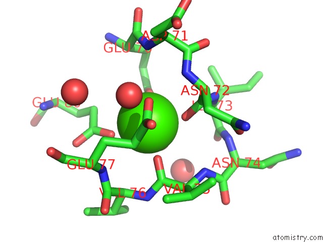
Mono view
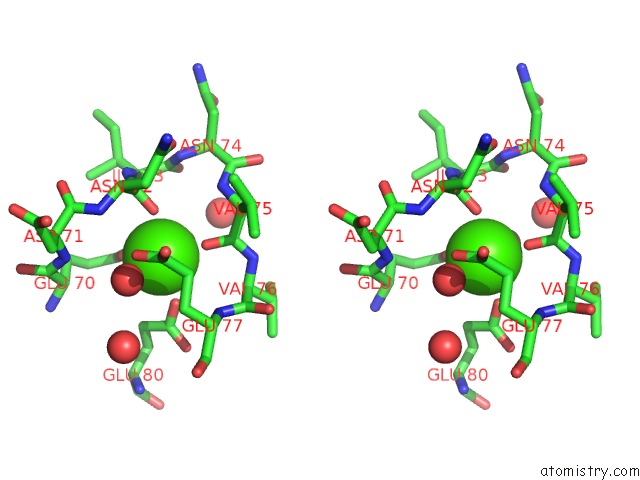
Stereo pair view
Mono view
Stereo pair view
A full contact list of Calcium with other atoms in the Ca binding
site number 1 of On the Disordered Activation Domain in Trypsinogen. Chemical Labelling and Low-Temperature Crystallography within 5.0Å range:
|
Reference:
J.Walter,
W.Steigemann,
T.P.Singh,
H.Bartunik,
W.Bode,
R.Huber.
On the Disordered Activation Domain in Trypsinogen. Chemical Labelling and Low-Temperature Crystallography Acta Crystallogr.,Sect.B V. 38 1462 1982.
ISSN: ISSN 0108-7681
Page generated: Sat Jul 13 16:56:38 2024
ISSN: ISSN 0108-7681
Last articles
Zn in 9J0NZn in 9J0O
Zn in 9J0P
Zn in 9FJX
Zn in 9EKB
Zn in 9C0F
Zn in 9CAH
Zn in 9CH0
Zn in 9CH3
Zn in 9CH1