Calcium »
PDB 3qgv-3r3t »
3qhq »
Calcium in PDB 3qhq: Structure of Crispr-Associated Protein CSN2
Enzymatic activity of Structure of Crispr-Associated Protein CSN2
All present enzymatic activity of Structure of Crispr-Associated Protein CSN2:
2.7.7.6;
2.7.7.6;
Protein crystallography data
The structure of Structure of Crispr-Associated Protein CSN2, PDB code: 3qhq
was solved by
P.Ellinger,
Z.Arslan,
R.Wurm,
B.Tschapek,
K.Pfeffer,
R.Wagner,
L.Schmitt,
U.Pul,
S.H.Smits,
with X-Ray Crystallography technique. A brief refinement statistics is given in the table below:
| Resolution Low / High (Å) | 19.57 / 2.00 |
| Space group | C 1 2 1 |
| Cell size a, b, c (Å), α, β, γ (°) | 75.300, 83.300, 110.400, 90.00, 109.40, 90.00 |
| R / Rfree (%) | 20.4 / 22.8 |
Calcium Binding Sites:
The binding sites of Calcium atom in the Structure of Crispr-Associated Protein CSN2
(pdb code 3qhq). This binding sites where shown within
5.0 Angstroms radius around Calcium atom.
In total 5 binding sites of Calcium where determined in the Structure of Crispr-Associated Protein CSN2, PDB code: 3qhq:
Jump to Calcium binding site number: 1; 2; 3; 4; 5;
In total 5 binding sites of Calcium where determined in the Structure of Crispr-Associated Protein CSN2, PDB code: 3qhq:
Jump to Calcium binding site number: 1; 2; 3; 4; 5;
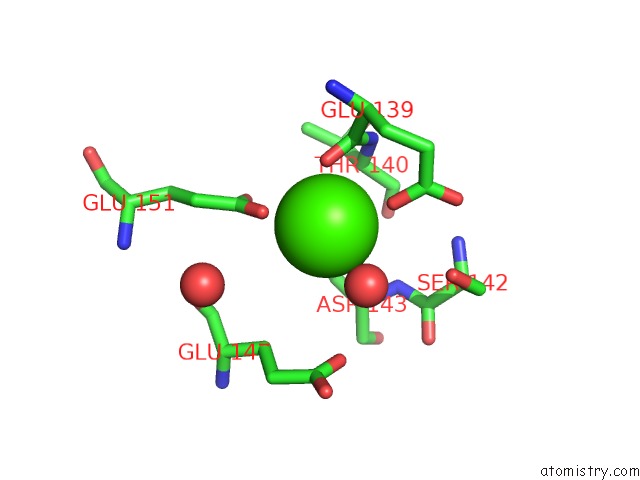
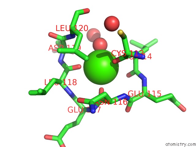
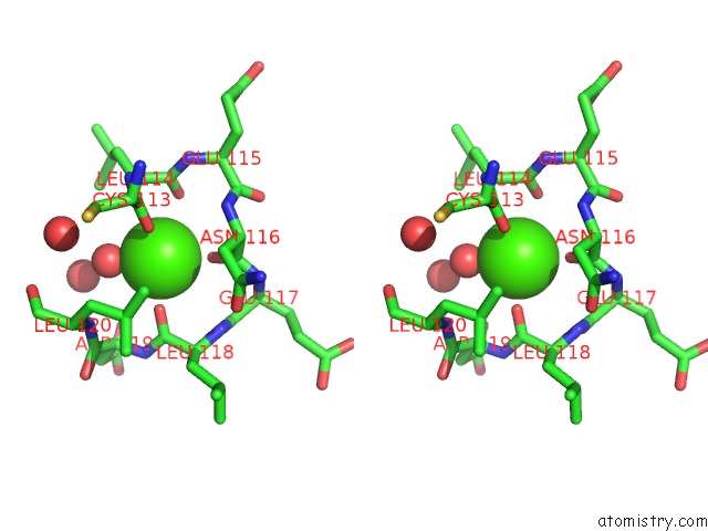
Calcium binding site 1 out of 5 in 3qhq
Go back to
Calcium binding site 1 out
of 5 in the Structure of Crispr-Associated Protein CSN2
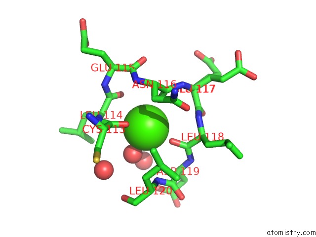
Mono view
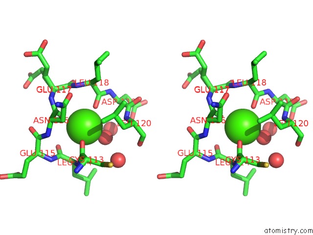
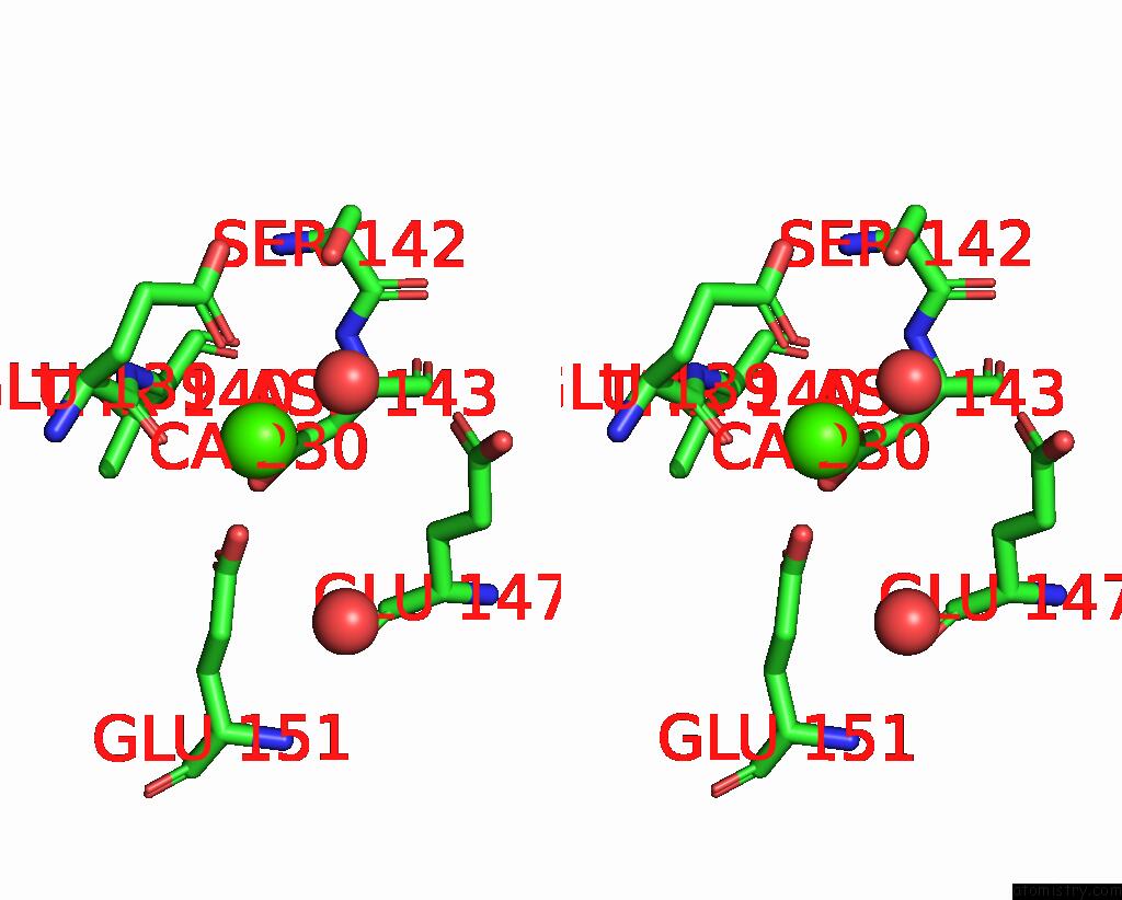
Stereo pair view
Mono view
Stereo pair view
A full contact list of Calcium with other atoms in the Ca binding
site number 1 of Structure of Crispr-Associated Protein CSN2 within 5.0Å range:
|
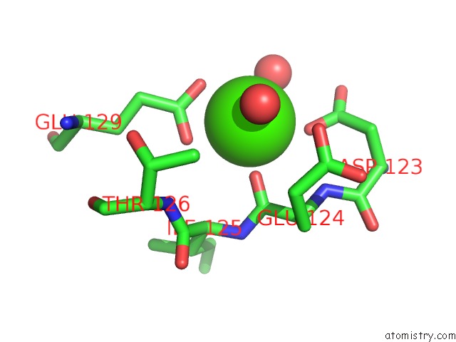
Calcium binding site 2 out of 5 in 3qhq
Go back to
Calcium binding site 2 out
of 5 in the Structure of Crispr-Associated Protein CSN2
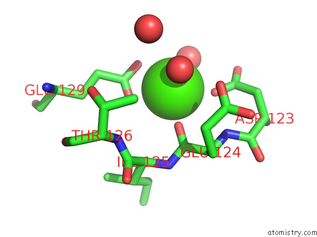
Mono view
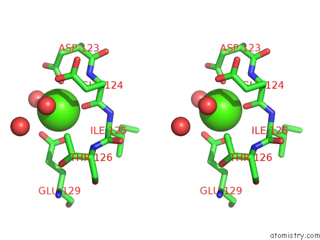
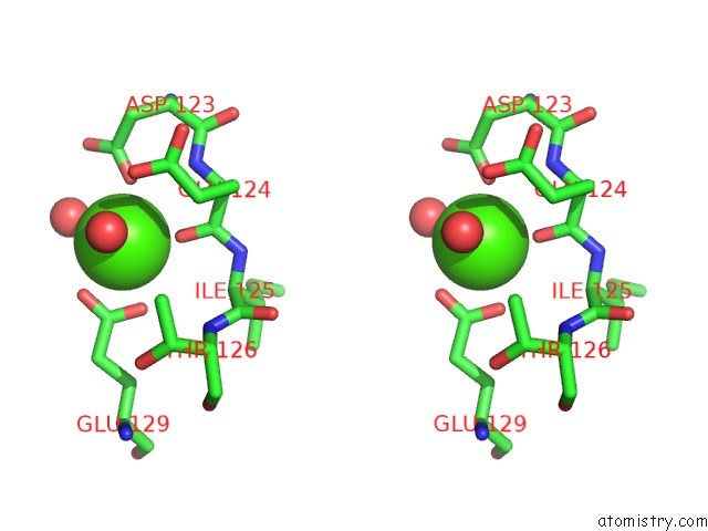
Stereo pair view
Mono view
Stereo pair view
A full contact list of Calcium with other atoms in the Ca binding
site number 2 of Structure of Crispr-Associated Protein CSN2 within 5.0Å range:
|
Calcium binding site 3 out of 5 in 3qhq
Go back to
Calcium binding site 3 out
of 5 in the Structure of Crispr-Associated Protein CSN2
Mono view
Stereo pair view
Mono view
Stereo pair view
A full contact list of Calcium with other atoms in the Ca binding
site number 3 of Structure of Crispr-Associated Protein CSN2 within 5.0Å range:
|
Calcium binding site 4 out of 5 in 3qhq
Go back to
Calcium binding site 4 out
of 5 in the Structure of Crispr-Associated Protein CSN2
Mono view
Stereo pair view
Mono view
Stereo pair view
A full contact list of Calcium with other atoms in the Ca binding
site number 4 of Structure of Crispr-Associated Protein CSN2 within 5.0Å range:
|
Calcium binding site 5 out of 5 in 3qhq
Go back to
Calcium binding site 5 out
of 5 in the Structure of Crispr-Associated Protein CSN2
Mono view
Stereo pair view
Mono view
Stereo pair view
A full contact list of Calcium with other atoms in the Ca binding
site number 5 of Structure of Crispr-Associated Protein CSN2 within 5.0Å range:
|
Reference:
P.Ellinger,
Z.Arslan,
R.Wurm,
B.Tschapek,
K.Pfeffer,
R.Wagner,
L.Schmitt,
U.Pul,
S.H.Smits.
Structure of Crispr-Associated Protein CSN2 To Be Published.
Page generated: Tue Jul 8 15:59:32 2025
Last articles
Ca in 7G3CCa in 7G3B
Ca in 7G3A
Ca in 7G39
Ca in 7G38
Ca in 7G37
Ca in 7G36
Ca in 7G33
Ca in 7G35
Ca in 7G32