Calcium »
PDB 3qgv-3r3t »
3r1h »
Calcium in PDB 3r1h: Crystal Structure of the Class I Ligase Ribozyme-Substrate Preligation Complex, C47U Mutant, CA2+ Bound
Protein crystallography data
The structure of Crystal Structure of the Class I Ligase Ribozyme-Substrate Preligation Complex, C47U Mutant, CA2+ Bound, PDB code: 3r1h
was solved by
D.M.Shechner,
D.P.Bartel,
with X-Ray Crystallography technique. A brief refinement statistics is given in the table below:
| Resolution Low / High (Å) | 30.00 / 3.15 |
| Space group | P 1 |
| Cell size a, b, c (Å), α, β, γ (°) | 58.690, 70.011, 71.858, 99.85, 99.74, 103.65 |
| R / Rfree (%) | 21.2 / 25.5 |
Calcium Binding Sites:
Pages:
>>> Page 1 <<< Page 2, Binding sites: 11 - 20; Page 3, Binding sites: 21 - 26;Binding sites:
The binding sites of Calcium atom in the Crystal Structure of the Class I Ligase Ribozyme-Substrate Preligation Complex, C47U Mutant, CA2+ Bound (pdb code 3r1h). This binding sites where shown within 5.0 Angstroms radius around Calcium atom.In total 26 binding sites of Calcium where determined in the Crystal Structure of the Class I Ligase Ribozyme-Substrate Preligation Complex, C47U Mutant, CA2+ Bound, PDB code: 3r1h:
Jump to Calcium binding site number: 1; 2; 3; 4; 5; 6; 7; 8; 9; 10;
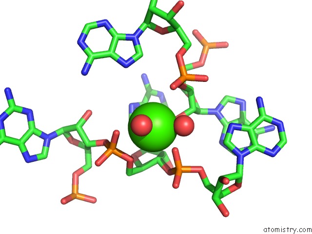
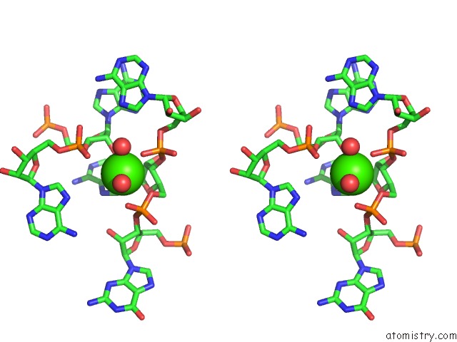
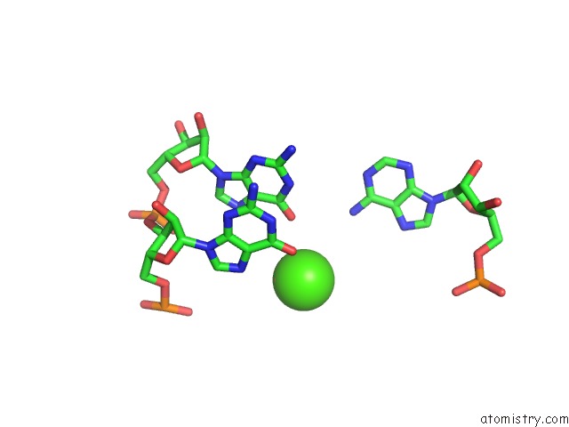
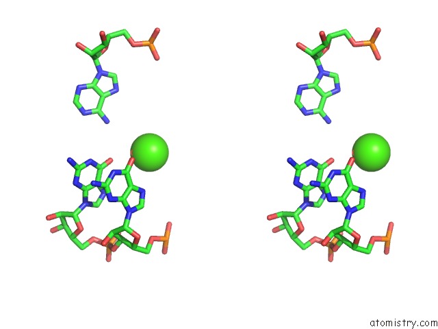
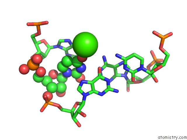
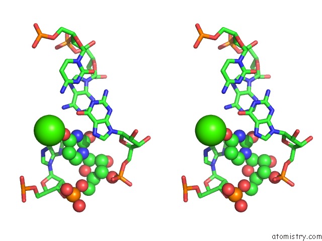
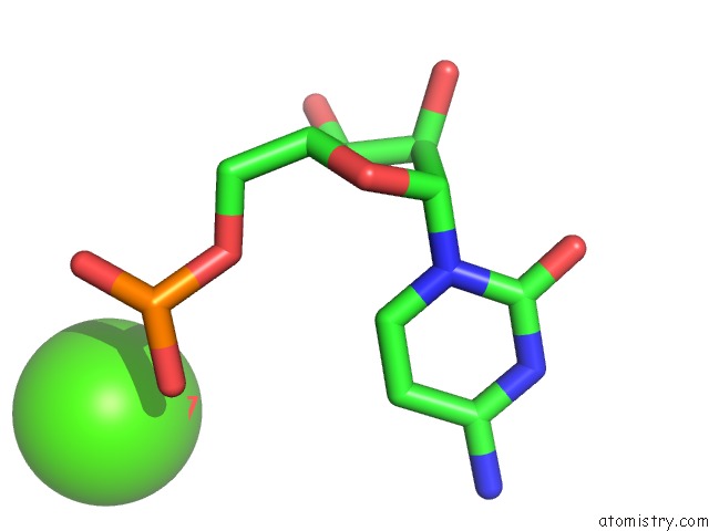
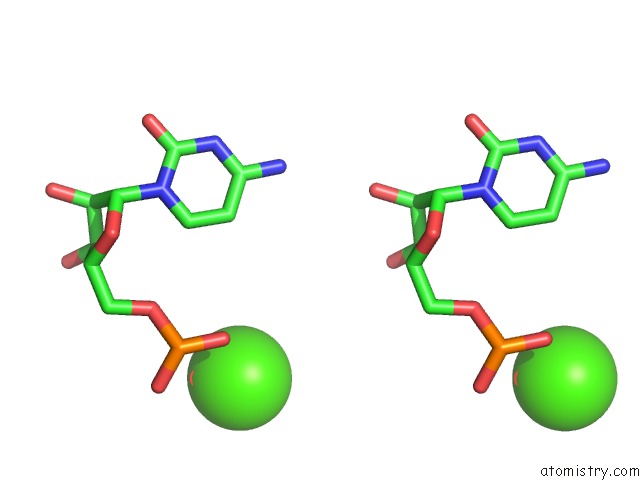
Calcium binding site 1 out of 26 in 3r1h
Go back to
Calcium binding site 1 out
of 26 in the Crystal Structure of the Class I Ligase Ribozyme-Substrate Preligation Complex, C47U Mutant, CA2+ Bound
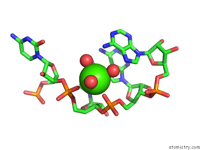
Mono view
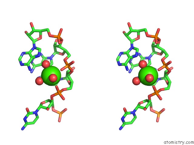
Stereo pair view
Mono view
Stereo pair view
A full contact list of Calcium with other atoms in the Ca binding
site number 1 of Crystal Structure of the Class I Ligase Ribozyme-Substrate Preligation Complex, C47U Mutant, CA2+ Bound within 5.0Å range:
|
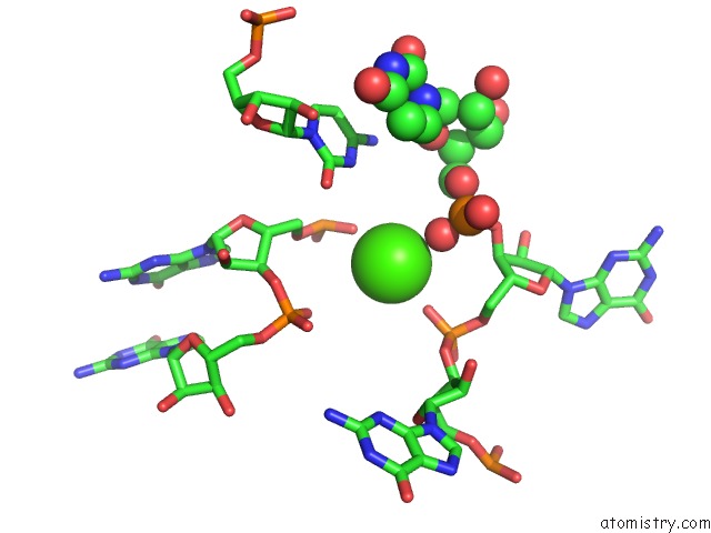
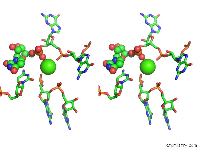
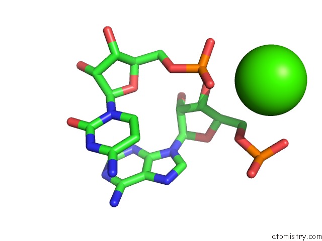
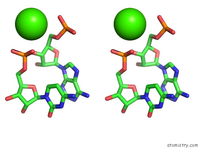
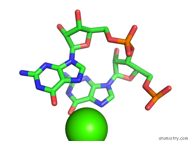
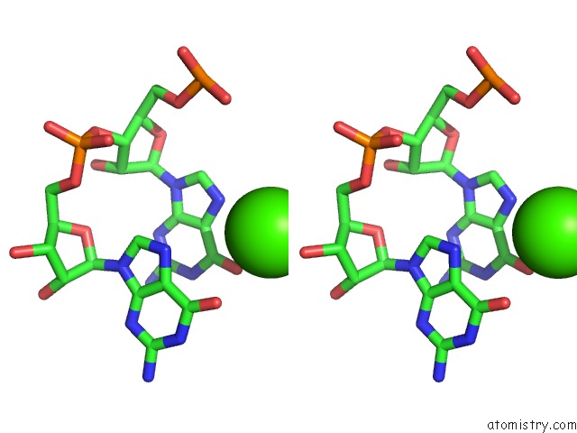
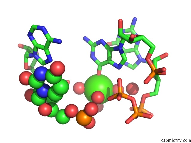
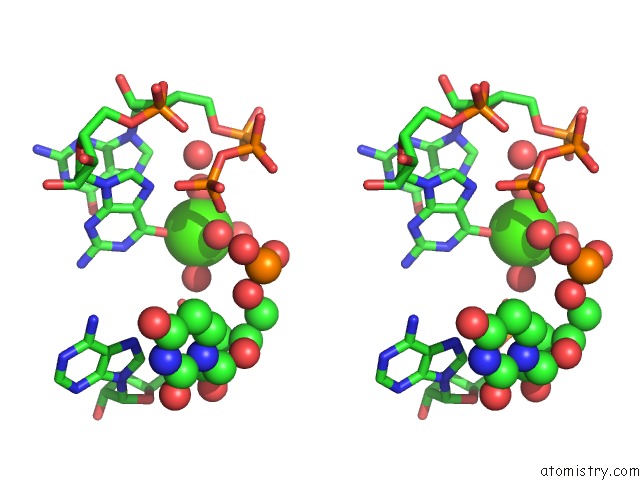
Calcium binding site 2 out of 26 in 3r1h
Go back to
Calcium binding site 2 out
of 26 in the Crystal Structure of the Class I Ligase Ribozyme-Substrate Preligation Complex, C47U Mutant, CA2+ Bound
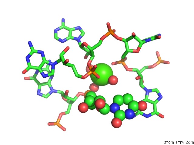
Mono view
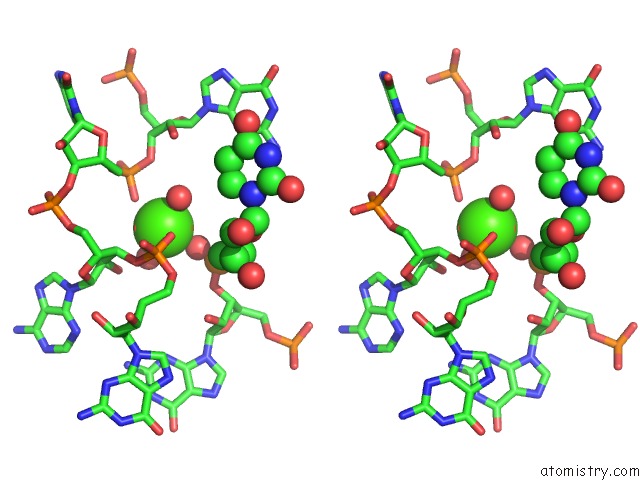
Stereo pair view
Mono view
Stereo pair view
A full contact list of Calcium with other atoms in the Ca binding
site number 2 of Crystal Structure of the Class I Ligase Ribozyme-Substrate Preligation Complex, C47U Mutant, CA2+ Bound within 5.0Å range:
|
Calcium binding site 3 out of 26 in 3r1h
Go back to
Calcium binding site 3 out
of 26 in the Crystal Structure of the Class I Ligase Ribozyme-Substrate Preligation Complex, C47U Mutant, CA2+ Bound
Mono view
Stereo pair view
Mono view
Stereo pair view
A full contact list of Calcium with other atoms in the Ca binding
site number 3 of Crystal Structure of the Class I Ligase Ribozyme-Substrate Preligation Complex, C47U Mutant, CA2+ Bound within 5.0Å range:
|
Calcium binding site 4 out of 26 in 3r1h
Go back to
Calcium binding site 4 out
of 26 in the Crystal Structure of the Class I Ligase Ribozyme-Substrate Preligation Complex, C47U Mutant, CA2+ Bound
Mono view
Stereo pair view
Mono view
Stereo pair view
A full contact list of Calcium with other atoms in the Ca binding
site number 4 of Crystal Structure of the Class I Ligase Ribozyme-Substrate Preligation Complex, C47U Mutant, CA2+ Bound within 5.0Å range:
|
Calcium binding site 5 out of 26 in 3r1h
Go back to
Calcium binding site 5 out
of 26 in the Crystal Structure of the Class I Ligase Ribozyme-Substrate Preligation Complex, C47U Mutant, CA2+ Bound
Mono view
Stereo pair view
Mono view
Stereo pair view
A full contact list of Calcium with other atoms in the Ca binding
site number 5 of Crystal Structure of the Class I Ligase Ribozyme-Substrate Preligation Complex, C47U Mutant, CA2+ Bound within 5.0Å range:
|
Calcium binding site 6 out of 26 in 3r1h
Go back to
Calcium binding site 6 out
of 26 in the Crystal Structure of the Class I Ligase Ribozyme-Substrate Preligation Complex, C47U Mutant, CA2+ Bound
Mono view
Stereo pair view
Mono view
Stereo pair view
A full contact list of Calcium with other atoms in the Ca binding
site number 6 of Crystal Structure of the Class I Ligase Ribozyme-Substrate Preligation Complex, C47U Mutant, CA2+ Bound within 5.0Å range:
|
Calcium binding site 7 out of 26 in 3r1h
Go back to
Calcium binding site 7 out
of 26 in the Crystal Structure of the Class I Ligase Ribozyme-Substrate Preligation Complex, C47U Mutant, CA2+ Bound
Mono view
Stereo pair view
Mono view
Stereo pair view
A full contact list of Calcium with other atoms in the Ca binding
site number 7 of Crystal Structure of the Class I Ligase Ribozyme-Substrate Preligation Complex, C47U Mutant, CA2+ Bound within 5.0Å range:
|
Calcium binding site 8 out of 26 in 3r1h
Go back to
Calcium binding site 8 out
of 26 in the Crystal Structure of the Class I Ligase Ribozyme-Substrate Preligation Complex, C47U Mutant, CA2+ Bound
Mono view
Stereo pair view
Mono view
Stereo pair view
A full contact list of Calcium with other atoms in the Ca binding
site number 8 of Crystal Structure of the Class I Ligase Ribozyme-Substrate Preligation Complex, C47U Mutant, CA2+ Bound within 5.0Å range:
|
Calcium binding site 9 out of 26 in 3r1h
Go back to
Calcium binding site 9 out
of 26 in the Crystal Structure of the Class I Ligase Ribozyme-Substrate Preligation Complex, C47U Mutant, CA2+ Bound
Mono view
Stereo pair view
Mono view
Stereo pair view
A full contact list of Calcium with other atoms in the Ca binding
site number 9 of Crystal Structure of the Class I Ligase Ribozyme-Substrate Preligation Complex, C47U Mutant, CA2+ Bound within 5.0Å range:
|
Calcium binding site 10 out of 26 in 3r1h
Go back to
Calcium binding site 10 out
of 26 in the Crystal Structure of the Class I Ligase Ribozyme-Substrate Preligation Complex, C47U Mutant, CA2+ Bound
Mono view
Stereo pair view
Mono view
Stereo pair view
A full contact list of Calcium with other atoms in the Ca binding
site number 10 of Crystal Structure of the Class I Ligase Ribozyme-Substrate Preligation Complex, C47U Mutant, CA2+ Bound within 5.0Å range:
|
Reference:
D.M.Shechner,
D.P.Bartel.
The Structural Basis of Rna-Catalyzed Rna Polymerization. Nat.Struct.Mol.Biol. V. 18 1036 2011.
ISSN: ISSN 1545-9993
PubMed: 21857665
DOI: 10.1038/NSMB.2107
Page generated: Sat Jul 13 17:44:24 2024
ISSN: ISSN 1545-9993
PubMed: 21857665
DOI: 10.1038/NSMB.2107
Last articles
Zn in 9MJ5Zn in 9HNW
Zn in 9G0L
Zn in 9FNE
Zn in 9DZN
Zn in 9E0I
Zn in 9D32
Zn in 9DAK
Zn in 8ZXC
Zn in 8ZUF