Calcium »
PDB 3qgv-3r3t »
3r3t »
Calcium in PDB 3r3t: Crystal Structure of 30S Ribosomal Protein S From Bacillus Anthracis
Protein crystallography data
The structure of Crystal Structure of 30S Ribosomal Protein S From Bacillus Anthracis, PDB code: 3r3t
was solved by
Y.Kim,
M.Zhou,
K.Kwon,
W.F.Anderson,
A.Joachimiak,
Center For Structuralgenomics Of Infectious Diseases (Csgid),
with X-Ray Crystallography technique. A brief refinement statistics is given in the table below:
| Resolution Low / High (Å) | 45.61 / 2.30 |
| Space group | P 31 2 1 |
| Cell size a, b, c (Å), α, β, γ (°) | 82.680, 82.680, 59.165, 90.00, 90.00, 120.00 |
| R / Rfree (%) | 20.8 / 23.2 |
Calcium Binding Sites:
The binding sites of Calcium atom in the Crystal Structure of 30S Ribosomal Protein S From Bacillus Anthracis
(pdb code 3r3t). This binding sites where shown within
5.0 Angstroms radius around Calcium atom.
In total 2 binding sites of Calcium where determined in the Crystal Structure of 30S Ribosomal Protein S From Bacillus Anthracis, PDB code: 3r3t:
Jump to Calcium binding site number: 1; 2;
In total 2 binding sites of Calcium where determined in the Crystal Structure of 30S Ribosomal Protein S From Bacillus Anthracis, PDB code: 3r3t:
Jump to Calcium binding site number: 1; 2;
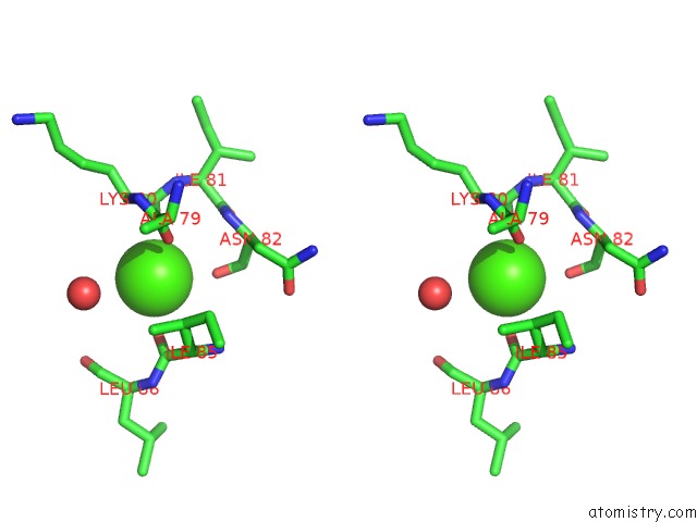
Calcium binding site 1 out of 2 in 3r3t
Go back to
Calcium binding site 1 out
of 2 in the Crystal Structure of 30S Ribosomal Protein S From Bacillus Anthracis

Mono view

Stereo pair view
Mono view
Stereo pair view
A full contact list of Calcium with other atoms in the Ca binding
site number 1 of Crystal Structure of 30S Ribosomal Protein S From Bacillus Anthracis within 5.0Å range:
|
Calcium binding site 2 out of 2 in 3r3t
Go back to
Calcium binding site 2 out
of 2 in the Crystal Structure of 30S Ribosomal Protein S From Bacillus Anthracis

Mono view
Stereo pair view
Mono view
Stereo pair view
A full contact list of Calcium with other atoms in the Ca binding
site number 2 of Crystal Structure of 30S Ribosomal Protein S From Bacillus Anthracis within 5.0Å range:
|
Reference:
Y.Kim,
M.Zhou,
K.Kwon,
W.F.Anderson,
A.Joachimiak,
Center For Structural Genomics Of Infectious Diseases(Csgid).
Crystal Structure of 30S Ribosomal Protein S From Bacillus Anthracis To Be Published.
Page generated: Tue Jul 8 16:08:02 2025
Last articles
F in 4HT0F in 4HNA
F in 4HPX
F in 4HQH
F in 4HNS
F in 4HPJ
F in 4HN4
F in 4HJX
F in 4HLH
F in 4HL4