Calcium »
PDB 3r3v-3rk2 »
3r68 »
Calcium in PDB 3r68: Molecular Analysis of the PDZ3 Domain of PDZK1
Protein crystallography data
The structure of Molecular Analysis of the PDZ3 Domain of PDZK1, PDB code: 3r68
was solved by
O.Kocher,
G.Birrane,
M.Krieger,
with X-Ray Crystallography technique. A brief refinement statistics is given in the table below:
| Resolution Low / High (Å) | 33.15 / 1.30 |
| Space group | P 43 21 2 |
| Cell size a, b, c (Å), α, β, γ (°) | 69.910, 69.910, 37.659, 90.00, 90.00, 90.00 |
| R / Rfree (%) | 13.1 / 15.7 |
Other elements in 3r68:
The structure of Molecular Analysis of the PDZ3 Domain of PDZK1 also contains other interesting chemical elements:
| Chlorine | (Cl) | 5 atoms |
| Zinc | (Zn) | 4 atoms |
Calcium Binding Sites:
The binding sites of Calcium atom in the Molecular Analysis of the PDZ3 Domain of PDZK1
(pdb code 3r68). This binding sites where shown within
5.0 Angstroms radius around Calcium atom.
In total 2 binding sites of Calcium where determined in the Molecular Analysis of the PDZ3 Domain of PDZK1, PDB code: 3r68:
Jump to Calcium binding site number: 1; 2;
In total 2 binding sites of Calcium where determined in the Molecular Analysis of the PDZ3 Domain of PDZK1, PDB code: 3r68:
Jump to Calcium binding site number: 1; 2;
Calcium binding site 1 out of 2 in 3r68
Go back to
Calcium binding site 1 out
of 2 in the Molecular Analysis of the PDZ3 Domain of PDZK1
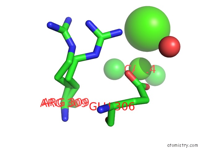
Mono view
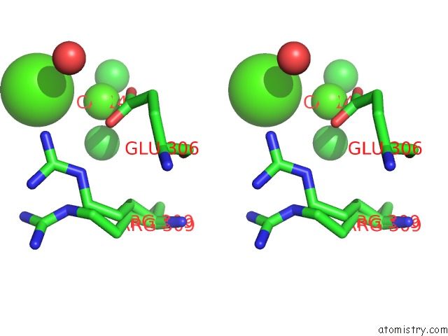
Stereo pair view
Mono view
Stereo pair view
A full contact list of Calcium with other atoms in the Ca binding
site number 1 of Molecular Analysis of the PDZ3 Domain of PDZK1 within 5.0Å range:
|
Calcium binding site 2 out of 2 in 3r68
Go back to
Calcium binding site 2 out
of 2 in the Molecular Analysis of the PDZ3 Domain of PDZK1
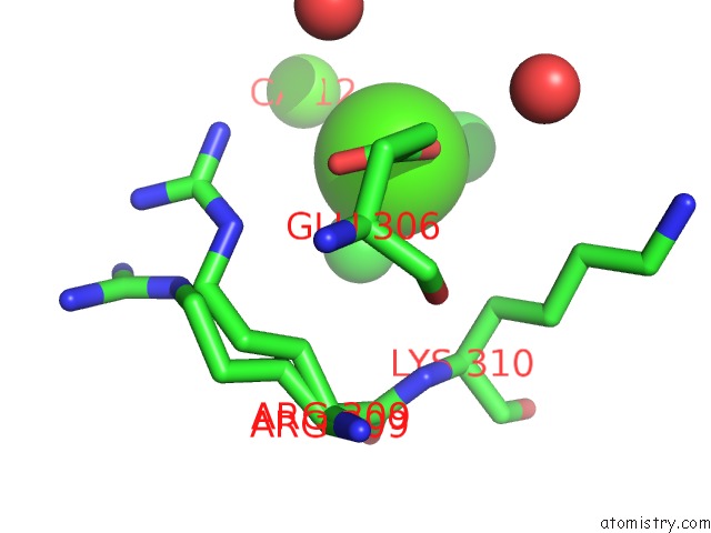
Mono view
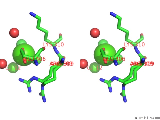
Stereo pair view
Mono view
Stereo pair view
A full contact list of Calcium with other atoms in the Ca binding
site number 2 of Molecular Analysis of the PDZ3 Domain of PDZK1 within 5.0Å range:
|
Reference:
O.Kocher,
G.Birrane,
A.Yesilaltay,
S.Shechter,
R.Pal,
K.Daniels,
M.Krieger.
Identification of the PDZ3 Domain of the Adaptor Protein PDZK1 As A Second, Physiologically Functional Binding Site For the C Terminus of the High Density Lipoprotein Receptor Scavenger Receptor Class B Type I. J.Biol.Chem. V. 286 25171 2011.
ISSN: ISSN 0021-9258
PubMed: 21602281
DOI: 10.1074/JBC.M111.242362
Page generated: Tue Jul 8 16:11:44 2025
ISSN: ISSN 0021-9258
PubMed: 21602281
DOI: 10.1074/JBC.M111.242362
Last articles
Cl in 5S4JCl in 5S33
Cl in 5S4I
Cl in 5RZ4
Cl in 5S2Y
Cl in 5S2W
Cl in 5S2U
Cl in 5S2N
Cl in 5RZO
Cl in 5RZG