Calcium »
PDB 3r3v-3rk2 »
3rbd »
Calcium in PDB 3rbd: DPO4 Extension Ternary Complex with 3'-Terminal Primer C Base Opposite the 3-Methylcytosine (M3C) Lesion
Enzymatic activity of DPO4 Extension Ternary Complex with 3'-Terminal Primer C Base Opposite the 3-Methylcytosine (M3C) Lesion
All present enzymatic activity of DPO4 Extension Ternary Complex with 3'-Terminal Primer C Base Opposite the 3-Methylcytosine (M3C) Lesion:
2.7.7.7;
2.7.7.7;
Protein crystallography data
The structure of DPO4 Extension Ternary Complex with 3'-Terminal Primer C Base Opposite the 3-Methylcytosine (M3C) Lesion, PDB code: 3rbd
was solved by
O.Rechkoblit,
D.J.Patel,
with X-Ray Crystallography technique. A brief refinement statistics is given in the table below:
| Resolution Low / High (Å) | 20.00 / 2.50 |
| Space group | P 1 21 1 |
| Cell size a, b, c (Å), α, β, γ (°) | 52.852, 109.339, 100.680, 90.00, 101.12, 90.00 |
| R / Rfree (%) | 20.4 / 23.8 |
Calcium Binding Sites:
The binding sites of Calcium atom in the DPO4 Extension Ternary Complex with 3'-Terminal Primer C Base Opposite the 3-Methylcytosine (M3C) Lesion
(pdb code 3rbd). This binding sites where shown within
5.0 Angstroms radius around Calcium atom.
In total 6 binding sites of Calcium where determined in the DPO4 Extension Ternary Complex with 3'-Terminal Primer C Base Opposite the 3-Methylcytosine (M3C) Lesion, PDB code: 3rbd:
Jump to Calcium binding site number: 1; 2; 3; 4; 5; 6;
In total 6 binding sites of Calcium where determined in the DPO4 Extension Ternary Complex with 3'-Terminal Primer C Base Opposite the 3-Methylcytosine (M3C) Lesion, PDB code: 3rbd:
Jump to Calcium binding site number: 1; 2; 3; 4; 5; 6;
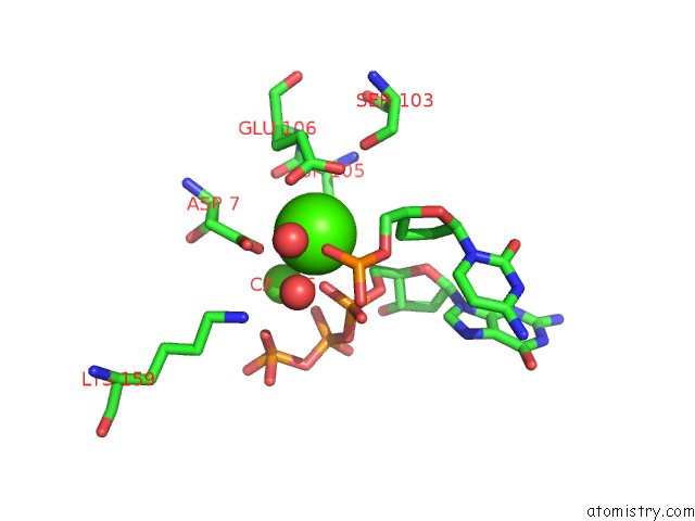
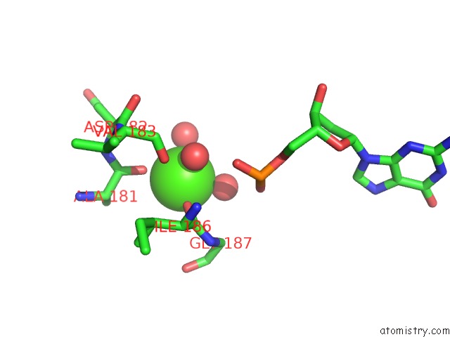
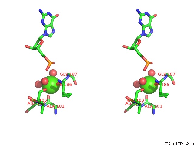
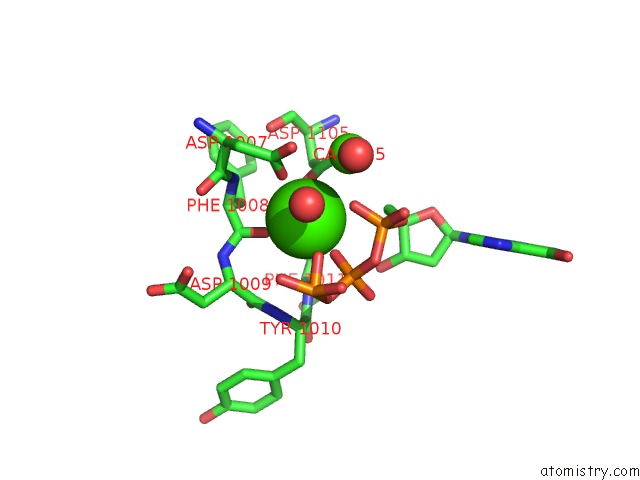
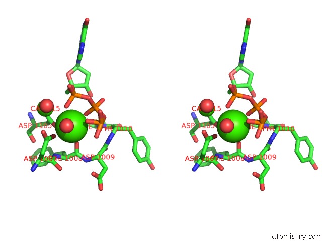
Calcium binding site 1 out of 6 in 3rbd
Go back to
Calcium binding site 1 out
of 6 in the DPO4 Extension Ternary Complex with 3'-Terminal Primer C Base Opposite the 3-Methylcytosine (M3C) Lesion
Mono view
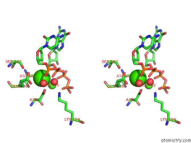
Stereo pair view
Mono view
Stereo pair view
A full contact list of Calcium with other atoms in the Ca binding
site number 1 of DPO4 Extension Ternary Complex with 3'-Terminal Primer C Base Opposite the 3-Methylcytosine (M3C) Lesion within 5.0Å range:
|
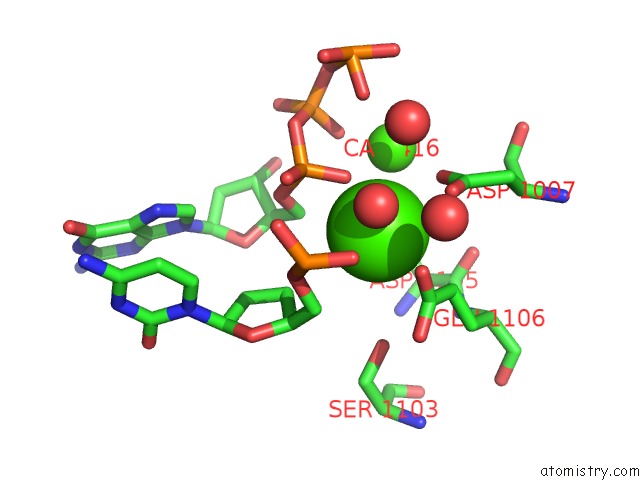
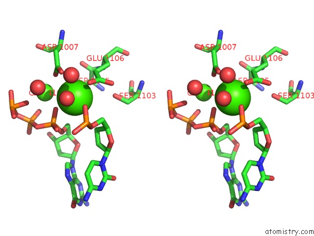
Calcium binding site 2 out of 6 in 3rbd
Go back to
Calcium binding site 2 out
of 6 in the DPO4 Extension Ternary Complex with 3'-Terminal Primer C Base Opposite the 3-Methylcytosine (M3C) Lesion
Mono view
Stereo pair view
Mono view
Stereo pair view
A full contact list of Calcium with other atoms in the Ca binding
site number 2 of DPO4 Extension Ternary Complex with 3'-Terminal Primer C Base Opposite the 3-Methylcytosine (M3C) Lesion within 5.0Å range:
|
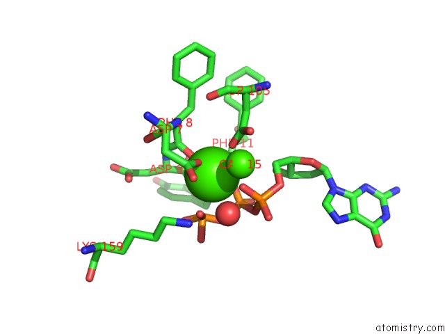
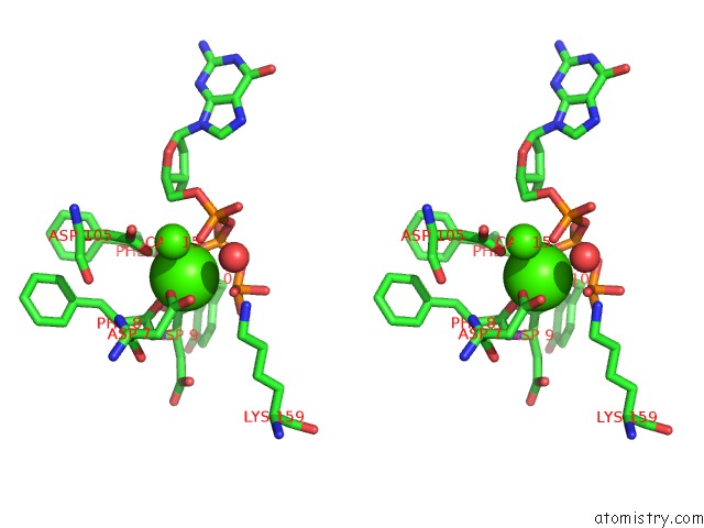
Calcium binding site 3 out of 6 in 3rbd
Go back to
Calcium binding site 3 out
of 6 in the DPO4 Extension Ternary Complex with 3'-Terminal Primer C Base Opposite the 3-Methylcytosine (M3C) Lesion
Mono view
Stereo pair view
Mono view
Stereo pair view
A full contact list of Calcium with other atoms in the Ca binding
site number 3 of DPO4 Extension Ternary Complex with 3'-Terminal Primer C Base Opposite the 3-Methylcytosine (M3C) Lesion within 5.0Å range:
|
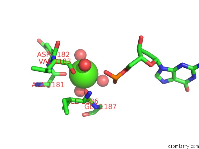
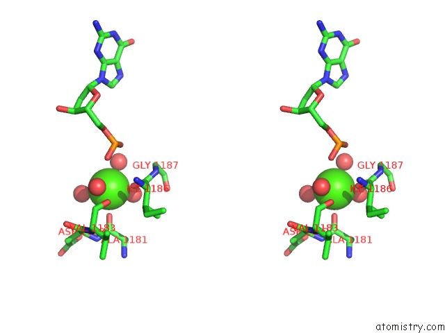
Calcium binding site 4 out of 6 in 3rbd
Go back to
Calcium binding site 4 out
of 6 in the DPO4 Extension Ternary Complex with 3'-Terminal Primer C Base Opposite the 3-Methylcytosine (M3C) Lesion
Mono view
Stereo pair view
Mono view
Stereo pair view
A full contact list of Calcium with other atoms in the Ca binding
site number 4 of DPO4 Extension Ternary Complex with 3'-Terminal Primer C Base Opposite the 3-Methylcytosine (M3C) Lesion within 5.0Å range:
|
Calcium binding site 5 out of 6 in 3rbd
Go back to
Calcium binding site 5 out
of 6 in the DPO4 Extension Ternary Complex with 3'-Terminal Primer C Base Opposite the 3-Methylcytosine (M3C) Lesion
Mono view
Stereo pair view
Mono view
Stereo pair view
A full contact list of Calcium with other atoms in the Ca binding
site number 5 of DPO4 Extension Ternary Complex with 3'-Terminal Primer C Base Opposite the 3-Methylcytosine (M3C) Lesion within 5.0Å range:
|
Calcium binding site 6 out of 6 in 3rbd
Go back to
Calcium binding site 6 out
of 6 in the DPO4 Extension Ternary Complex with 3'-Terminal Primer C Base Opposite the 3-Methylcytosine (M3C) Lesion
Mono view
Stereo pair view
Mono view
Stereo pair view
A full contact list of Calcium with other atoms in the Ca binding
site number 6 of DPO4 Extension Ternary Complex with 3'-Terminal Primer C Base Opposite the 3-Methylcytosine (M3C) Lesion within 5.0Å range:
|
Reference:
O.Rechkoblit,
J.C.Delaney,
J.M.Essigmann,
D.J.Patel.
Implications For Damage Recognition During DPO4-Mediated Mutagenic Bypass of M1G and M3C Lesions. Structure V. 19 821 2011.
ISSN: ISSN 0969-2126
PubMed: 21645853
DOI: 10.1016/J.STR.2011.03.020
Page generated: Sat Jul 13 17:59:08 2024
ISSN: ISSN 0969-2126
PubMed: 21645853
DOI: 10.1016/J.STR.2011.03.020
Last articles
Zn in 9J0NZn in 9J0O
Zn in 9J0P
Zn in 9FJX
Zn in 9EKB
Zn in 9C0F
Zn in 9CAH
Zn in 9CH0
Zn in 9CH3
Zn in 9CH1