Calcium »
PDB 3r3v-3rk2 »
3rk2 »
Calcium in PDB 3rk2: Truncated Snare Complex
Protein crystallography data
The structure of Truncated Snare Complex, PDB code: 3rk2
was solved by
D.Kuemmel,
K.M.Reinisch,
with X-Ray Crystallography technique. A brief refinement statistics is given in the table below:
| Resolution Low / High (Å) | 39.50 / 2.20 |
| Space group | P 1 |
| Cell size a, b, c (Å), α, β, γ (°) | 27.627, 39.769, 102.275, 83.38, 89.94, 89.87 |
| R / Rfree (%) | 22.7 / 26.8 |
Calcium Binding Sites:
The binding sites of Calcium atom in the Truncated Snare Complex
(pdb code 3rk2). This binding sites where shown within
5.0 Angstroms radius around Calcium atom.
In total 4 binding sites of Calcium where determined in the Truncated Snare Complex, PDB code: 3rk2:
Jump to Calcium binding site number: 1; 2; 3; 4;
In total 4 binding sites of Calcium where determined in the Truncated Snare Complex, PDB code: 3rk2:
Jump to Calcium binding site number: 1; 2; 3; 4;
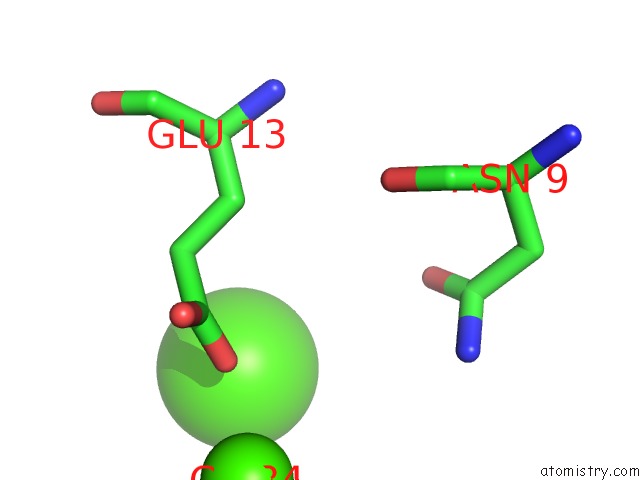
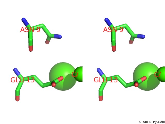
Calcium binding site 1 out of 4 in 3rk2
Go back to
Calcium binding site 1 out
of 4 in the Truncated Snare Complex
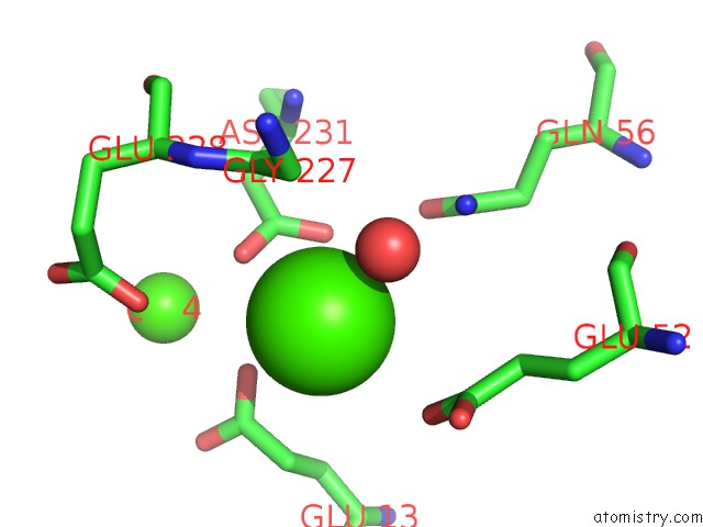
Mono view
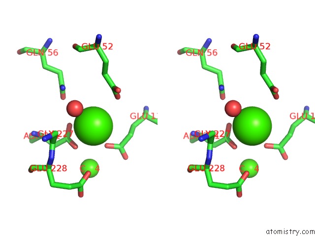
Stereo pair view
Mono view
Stereo pair view
A full contact list of Calcium with other atoms in the Ca binding
site number 1 of Truncated Snare Complex within 5.0Å range:
|
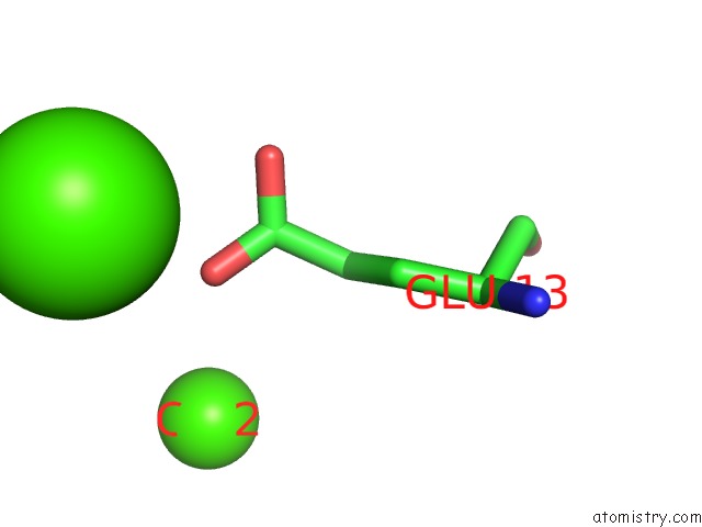
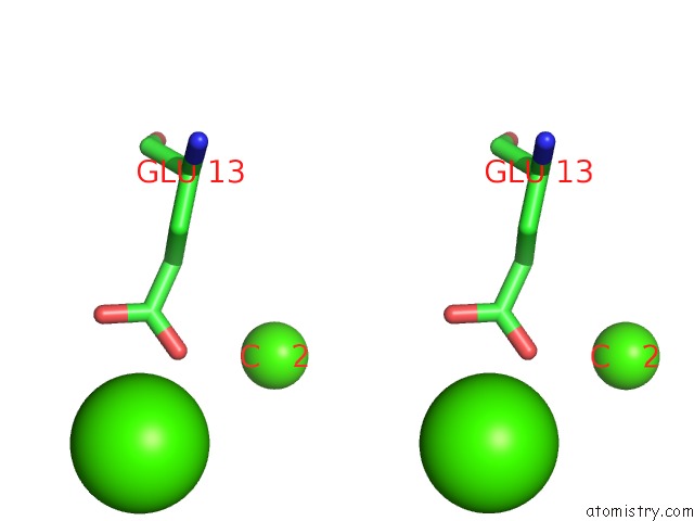
Calcium binding site 2 out of 4 in 3rk2
Go back to
Calcium binding site 2 out
of 4 in the Truncated Snare Complex
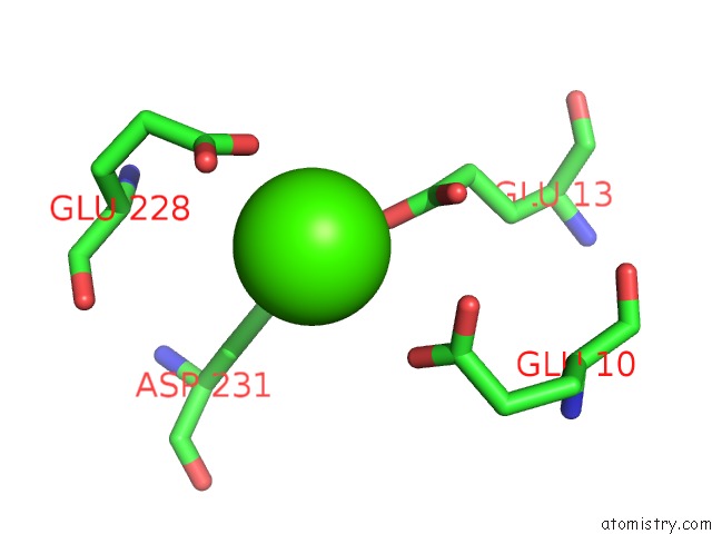
Mono view
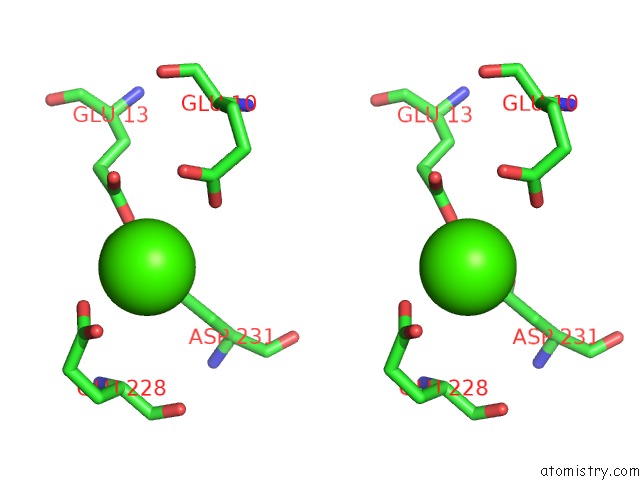
Stereo pair view
Mono view
Stereo pair view
A full contact list of Calcium with other atoms in the Ca binding
site number 2 of Truncated Snare Complex within 5.0Å range:
|
Calcium binding site 3 out of 4 in 3rk2
Go back to
Calcium binding site 3 out
of 4 in the Truncated Snare Complex
Mono view
Stereo pair view
Mono view
Stereo pair view
A full contact list of Calcium with other atoms in the Ca binding
site number 3 of Truncated Snare Complex within 5.0Å range:
|
Calcium binding site 4 out of 4 in 3rk2
Go back to
Calcium binding site 4 out
of 4 in the Truncated Snare Complex
Mono view
Stereo pair view
Mono view
Stereo pair view
A full contact list of Calcium with other atoms in the Ca binding
site number 4 of Truncated Snare Complex within 5.0Å range:
|
Reference:
D.Kummel,
S.S.Krishnakumar,
D.T.Radoff,
F.Li,
C.G.Giraudo,
F.Pincet,
J.E.Rothman,
K.M.Reinisch.
Complexin Cross-Links Prefusion Snares Into A Zigzag Array. Nat.Struct.Mol.Biol. V. 18 927 2011.
ISSN: ISSN 1545-9993
PubMed: 21785414
DOI: 10.1038/NSMB.2101
Page generated: Sat Jul 13 18:04:41 2024
ISSN: ISSN 1545-9993
PubMed: 21785414
DOI: 10.1038/NSMB.2101
Last articles
Zn in 9MJ5Zn in 9HNW
Zn in 9G0L
Zn in 9FNE
Zn in 9DZN
Zn in 9E0I
Zn in 9D32
Zn in 9DAK
Zn in 8ZXC
Zn in 8ZUF