Calcium »
PDB 3rke-3rxa »
3rup »
Calcium in PDB 3rup: Crystal Structure of E.Coli Biotin Carboxylase in Complex with Two Adp and Two Ca Ions
Enzymatic activity of Crystal Structure of E.Coli Biotin Carboxylase in Complex with Two Adp and Two Ca Ions
All present enzymatic activity of Crystal Structure of E.Coli Biotin Carboxylase in Complex with Two Adp and Two Ca Ions:
6.3.4.14; 6.4.1.2;
6.3.4.14; 6.4.1.2;
Protein crystallography data
The structure of Crystal Structure of E.Coli Biotin Carboxylase in Complex with Two Adp and Two Ca Ions, PDB code: 3rup
was solved by
C.Y.Chou,
L.Tong,
with X-Ray Crystallography technique. A brief refinement statistics is given in the table below:
| Resolution Low / High (Å) | 30.00 / 1.99 |
| Space group | C 1 2 1 |
| Cell size a, b, c (Å), α, β, γ (°) | 170.180, 58.843, 85.083, 90.00, 94.24, 90.00 |
| R / Rfree (%) | 17.1 / 22.7 |
Other elements in 3rup:
The structure of Crystal Structure of E.Coli Biotin Carboxylase in Complex with Two Adp and Two Ca Ions also contains other interesting chemical elements:
| Chlorine | (Cl) | 3 atoms |
Calcium Binding Sites:
The binding sites of Calcium atom in the Crystal Structure of E.Coli Biotin Carboxylase in Complex with Two Adp and Two Ca Ions
(pdb code 3rup). This binding sites where shown within
5.0 Angstroms radius around Calcium atom.
In total 4 binding sites of Calcium where determined in the Crystal Structure of E.Coli Biotin Carboxylase in Complex with Two Adp and Two Ca Ions, PDB code: 3rup:
Jump to Calcium binding site number: 1; 2; 3; 4;
In total 4 binding sites of Calcium where determined in the Crystal Structure of E.Coli Biotin Carboxylase in Complex with Two Adp and Two Ca Ions, PDB code: 3rup:
Jump to Calcium binding site number: 1; 2; 3; 4;
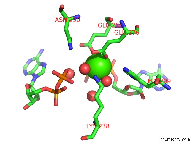
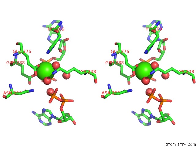
Calcium binding site 1 out of 4 in 3rup
Go back to
Calcium binding site 1 out
of 4 in the Crystal Structure of E.Coli Biotin Carboxylase in Complex with Two Adp and Two Ca Ions
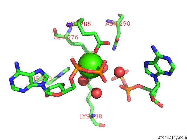
Mono view
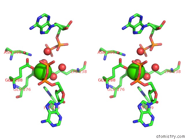
Stereo pair view
Mono view
Stereo pair view
A full contact list of Calcium with other atoms in the Ca binding
site number 1 of Crystal Structure of E.Coli Biotin Carboxylase in Complex with Two Adp and Two Ca Ions within 5.0Å range:
|
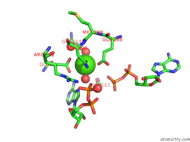
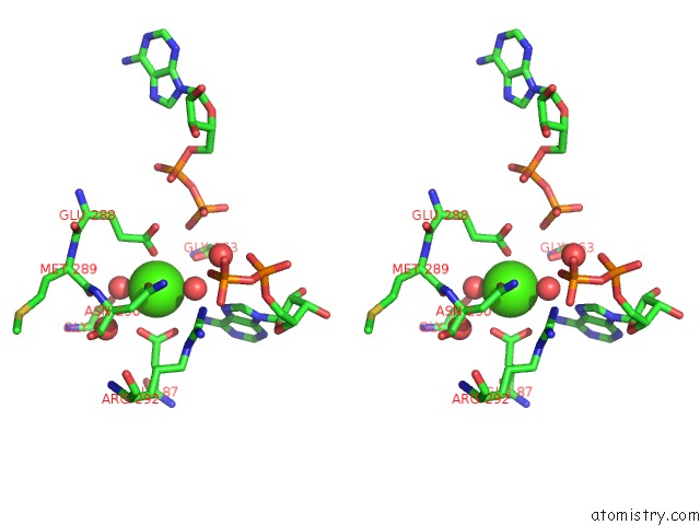
Calcium binding site 2 out of 4 in 3rup
Go back to
Calcium binding site 2 out
of 4 in the Crystal Structure of E.Coli Biotin Carboxylase in Complex with Two Adp and Two Ca Ions
Mono view
Stereo pair view
Mono view
Stereo pair view
A full contact list of Calcium with other atoms in the Ca binding
site number 2 of Crystal Structure of E.Coli Biotin Carboxylase in Complex with Two Adp and Two Ca Ions within 5.0Å range:
|
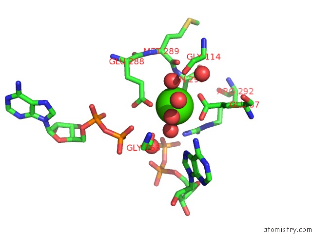
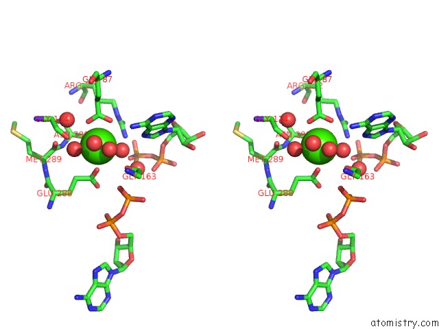
Calcium binding site 3 out of 4 in 3rup
Go back to
Calcium binding site 3 out
of 4 in the Crystal Structure of E.Coli Biotin Carboxylase in Complex with Two Adp and Two Ca Ions
Mono view
Stereo pair view
Mono view
Stereo pair view
A full contact list of Calcium with other atoms in the Ca binding
site number 3 of Crystal Structure of E.Coli Biotin Carboxylase in Complex with Two Adp and Two Ca Ions within 5.0Å range:
|
Calcium binding site 4 out of 4 in 3rup
Go back to
Calcium binding site 4 out
of 4 in the Crystal Structure of E.Coli Biotin Carboxylase in Complex with Two Adp and Two Ca Ions
Mono view
Stereo pair view
Mono view
Stereo pair view
A full contact list of Calcium with other atoms in the Ca binding
site number 4 of Crystal Structure of E.Coli Biotin Carboxylase in Complex with Two Adp and Two Ca Ions within 5.0Å range:
|
Reference:
C.Y.Chou,
L.Tong.
Structural and Biochemical Studies on the Regulation of Biotin Carboxylase By Substrate Inhibition and Dimerization. J.Biol.Chem. V. 286 24417 2011.
ISSN: ISSN 0021-9258
PubMed: 21592965
DOI: 10.1074/JBC.M111.220517
Page generated: Sat Jul 13 18:13:05 2024
ISSN: ISSN 0021-9258
PubMed: 21592965
DOI: 10.1074/JBC.M111.220517
Last articles
Zn in 9J0NZn in 9J0O
Zn in 9J0P
Zn in 9FJX
Zn in 9EKB
Zn in 9C0F
Zn in 9CAH
Zn in 9CH0
Zn in 9CH3
Zn in 9CH1