Calcium »
PDB 3rxb-3s9n »
3rxh »
Calcium in PDB 3rxh: Crystal Structure of Trypsin Complexed with 2-(1H-Imidazol-4-Yl) Ethanamine
Enzymatic activity of Crystal Structure of Trypsin Complexed with 2-(1H-Imidazol-4-Yl) Ethanamine
All present enzymatic activity of Crystal Structure of Trypsin Complexed with 2-(1H-Imidazol-4-Yl) Ethanamine:
3.4.21.4;
3.4.21.4;
Protein crystallography data
The structure of Crystal Structure of Trypsin Complexed with 2-(1H-Imidazol-4-Yl) Ethanamine, PDB code: 3rxh
was solved by
J.Yamane,
M.Yao,
Y.Zhou,
I.Tanaka,
with X-Ray Crystallography technique. A brief refinement statistics is given in the table below:
| Resolution Low / High (Å) | 20.00 / 1.70 |
| Space group | P 21 21 21 |
| Cell size a, b, c (Å), α, β, γ (°) | 54.401, 58.397, 66.646, 90.00, 90.00, 90.00 |
| R / Rfree (%) | 16.2 / 18.4 |
Calcium Binding Sites:
The binding sites of Calcium atom in the Crystal Structure of Trypsin Complexed with 2-(1H-Imidazol-4-Yl) Ethanamine
(pdb code 3rxh). This binding sites where shown within
5.0 Angstroms radius around Calcium atom.
In total only one binding site of Calcium was determined in the Crystal Structure of Trypsin Complexed with 2-(1H-Imidazol-4-Yl) Ethanamine, PDB code: 3rxh:
In total only one binding site of Calcium was determined in the Crystal Structure of Trypsin Complexed with 2-(1H-Imidazol-4-Yl) Ethanamine, PDB code: 3rxh:
Calcium binding site 1 out of 1 in 3rxh
Go back to
Calcium binding site 1 out
of 1 in the Crystal Structure of Trypsin Complexed with 2-(1H-Imidazol-4-Yl) Ethanamine
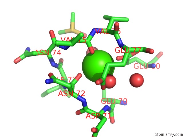
Mono view
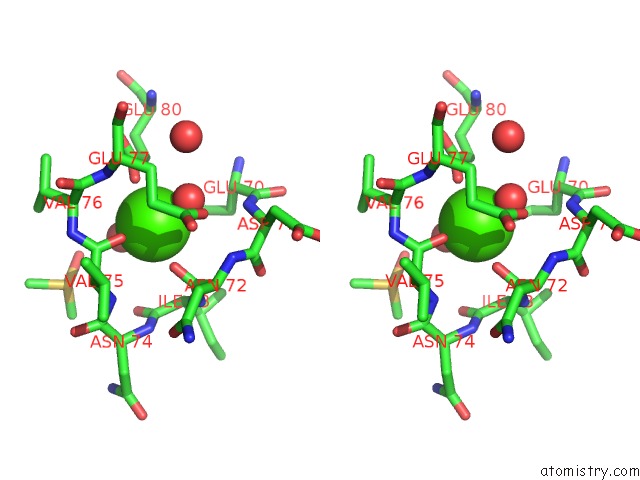
Stereo pair view
Mono view
Stereo pair view
A full contact list of Calcium with other atoms in the Ca binding
site number 1 of Crystal Structure of Trypsin Complexed with 2-(1H-Imidazol-4-Yl) Ethanamine within 5.0Å range:
|
Reference:
J.Yamane,
M.Yao,
Y.Zhou,
Y.Hiramatsu,
K.Fujiwara,
T.Yamaguchi,
H.Yamaguchi,
H.Togame,
H.Tsujishita,
H.Takemoto,
I.Tanaka.
In-Crystal Affinity Ranking of Fragment Hit Compounds Reveals A Relationship with Their Inhibitory Activities J.Appl.Crystallogr. V. 44 798 2011.
ISSN: ISSN 0021-8898
DOI: 10.1107/S0021889811017717
Page generated: Tue Jul 8 16:27:37 2025
ISSN: ISSN 0021-8898
DOI: 10.1107/S0021889811017717
Last articles
F in 7MYYF in 7N13
F in 7MYU
F in 7MYR
F in 7MYO
F in 7MXN
F in 7MXG
F in 7MXH
F in 7MX7
F in 7MVS