Calcium »
PDB 3rxb-3s9n »
3s82 »
Calcium in PDB 3s82: Structure of A S-Adenosylmethionine Synthetase From Mycobacterium Avium
Enzymatic activity of Structure of A S-Adenosylmethionine Synthetase From Mycobacterium Avium
All present enzymatic activity of Structure of A S-Adenosylmethionine Synthetase From Mycobacterium Avium:
2.5.1.6;
2.5.1.6;
Protein crystallography data
The structure of Structure of A S-Adenosylmethionine Synthetase From Mycobacterium Avium, PDB code: 3s82
was solved by
Seattle Structural Genomics Center For Infectious Disease,
Seattlestructural Genomics Center For Infectious Disease (Ssgcid),
with X-Ray Crystallography technique. A brief refinement statistics is given in the table below:
| Resolution Low / High (Å) | 20.00 / 1.73 |
| Space group | P 2 21 21 |
| Cell size a, b, c (Å), α, β, γ (°) | 60.030, 88.180, 184.190, 90.00, 90.00, 90.00 |
| R / Rfree (%) | 15.7 / 17.7 |
Other elements in 3s82:
The structure of Structure of A S-Adenosylmethionine Synthetase From Mycobacterium Avium also contains other interesting chemical elements:
| Uranium | (U) | 2 atoms |
Calcium Binding Sites:
The binding sites of Calcium atom in the Structure of A S-Adenosylmethionine Synthetase From Mycobacterium Avium
(pdb code 3s82). This binding sites where shown within
5.0 Angstroms radius around Calcium atom.
In total 4 binding sites of Calcium where determined in the Structure of A S-Adenosylmethionine Synthetase From Mycobacterium Avium, PDB code: 3s82:
Jump to Calcium binding site number: 1; 2; 3; 4;
In total 4 binding sites of Calcium where determined in the Structure of A S-Adenosylmethionine Synthetase From Mycobacterium Avium, PDB code: 3s82:
Jump to Calcium binding site number: 1; 2; 3; 4;
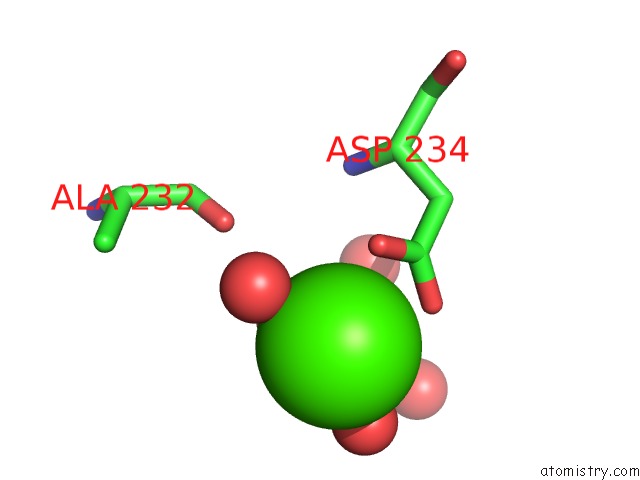
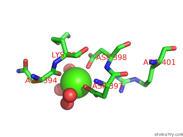
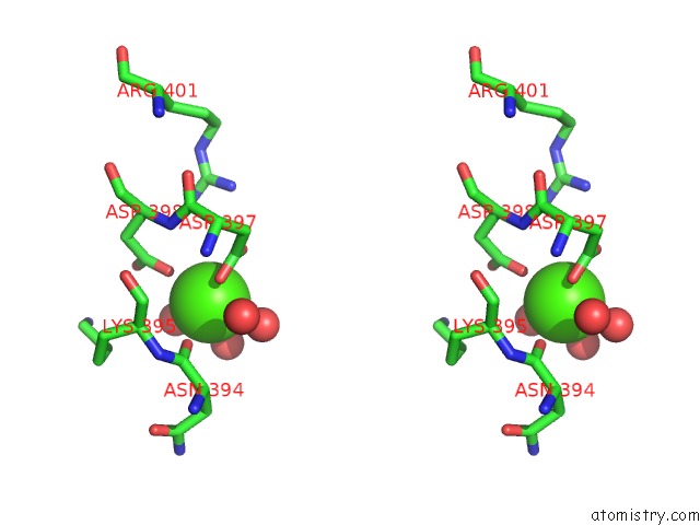
Calcium binding site 1 out of 4 in 3s82
Go back to
Calcium binding site 1 out
of 4 in the Structure of A S-Adenosylmethionine Synthetase From Mycobacterium Avium
Mono view
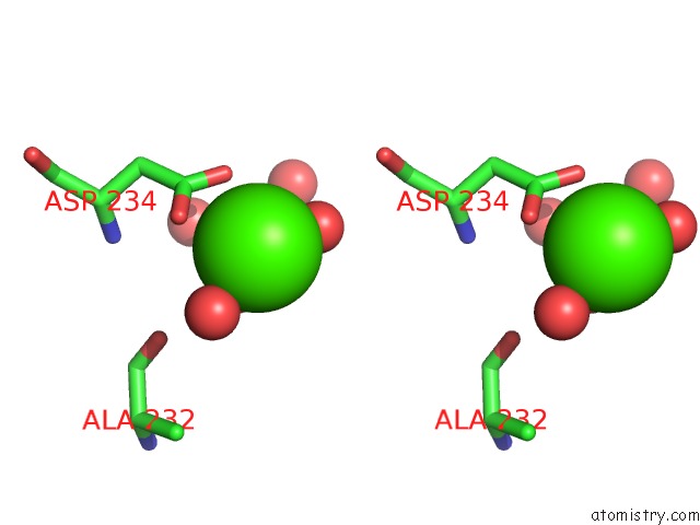
Stereo pair view
Mono view
Stereo pair view
A full contact list of Calcium with other atoms in the Ca binding
site number 1 of Structure of A S-Adenosylmethionine Synthetase From Mycobacterium Avium within 5.0Å range:
|
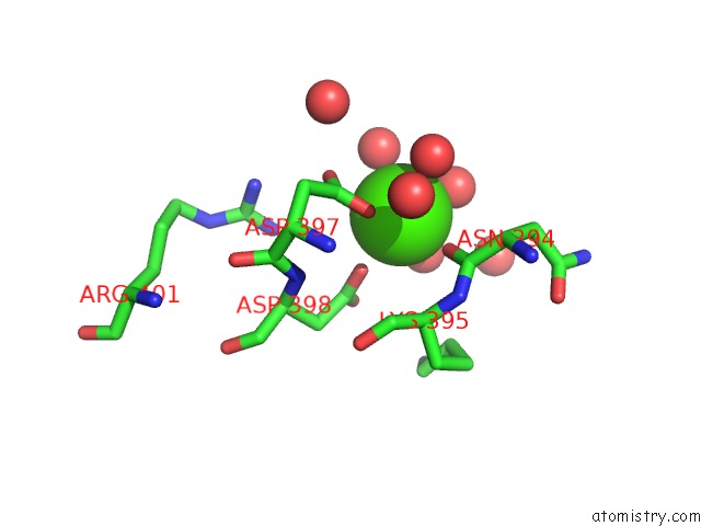
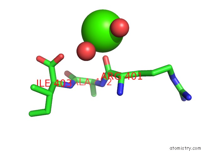
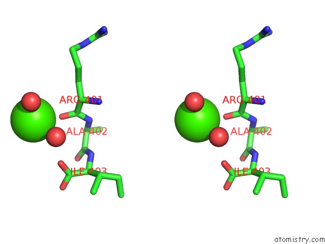
Calcium binding site 2 out of 4 in 3s82
Go back to
Calcium binding site 2 out
of 4 in the Structure of A S-Adenosylmethionine Synthetase From Mycobacterium Avium
Mono view
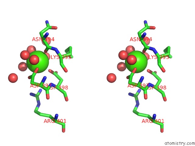
Stereo pair view
Mono view
Stereo pair view
A full contact list of Calcium with other atoms in the Ca binding
site number 2 of Structure of A S-Adenosylmethionine Synthetase From Mycobacterium Avium within 5.0Å range:
|
Calcium binding site 3 out of 4 in 3s82
Go back to
Calcium binding site 3 out
of 4 in the Structure of A S-Adenosylmethionine Synthetase From Mycobacterium Avium
Mono view
Stereo pair view
Mono view
Stereo pair view
A full contact list of Calcium with other atoms in the Ca binding
site number 3 of Structure of A S-Adenosylmethionine Synthetase From Mycobacterium Avium within 5.0Å range:
|
Calcium binding site 4 out of 4 in 3s82
Go back to
Calcium binding site 4 out
of 4 in the Structure of A S-Adenosylmethionine Synthetase From Mycobacterium Avium
Mono view
Stereo pair view
Mono view
Stereo pair view
A full contact list of Calcium with other atoms in the Ca binding
site number 4 of Structure of A S-Adenosylmethionine Synthetase From Mycobacterium Avium within 5.0Å range:
|
Reference:
L.Baugh,
I.Phan,
D.W.Begley,
M.C.Clifton,
B.Armour,
D.M.Dranow,
B.M.Taylor,
M.M.Muruthi,
J.Abendroth,
J.W.Fairman,
D.Fox,
S.H.Dieterich,
B.L.Staker,
A.S.Gardberg,
R.Choi,
S.N.Hewitt,
A.J.Napuli,
J.Myers,
L.K.Barrett,
Y.Zhang,
M.Ferrell,
E.Mundt,
K.Thompkins,
N.Tran,
S.Lyons-Abbott,
A.Abramov,
A.Sekar,
D.Serbzhinskiy,
D.Lorimer,
G.W.Buchko,
R.Stacy,
L.J.Stewart,
T.E.Edwards,
W.C.Van Voorhis,
P.J.Myler.
Increasing the Structural Coverage of Tuberculosis Drug Targets. Tuberculosis (Edinb) V. 95 142 2015.
ISSN: ISSN 1472-9792
PubMed: 25613812
DOI: 10.1016/J.TUBE.2014.12.003
Page generated: Tue Jul 8 16:31:48 2025
ISSN: ISSN 1472-9792
PubMed: 25613812
DOI: 10.1016/J.TUBE.2014.12.003
Last articles
Ca in 7LQBCa in 7LQA
Ca in 7LRF
Ca in 7LQ9
Ca in 7LQ8
Ca in 7LPV
Ca in 7LPU
Ca in 7LO5
Ca in 7LNP
Ca in 7LPT