Calcium »
PDB 3s9w-3snz »
3sny »
Calcium in PDB 3sny: Crystal Structure of A Mutant T82R of A Betagamma-Crystallin Domain From Clostridium Beijerinckii
Protein crystallography data
The structure of Crystal Structure of A Mutant T82R of A Betagamma-Crystallin Domain From Clostridium Beijerinckii, PDB code: 3sny
was solved by
S.S.Srivastava,
R.Sankaranarayanan,
with X-Ray Crystallography technique. A brief refinement statistics is given in the table below:
| Resolution Low / High (Å) | 50.00 / 1.85 |
| Space group | I 4 2 2 |
| Cell size a, b, c (Å), α, β, γ (°) | 77.314, 77.314, 77.738, 90.00, 90.00, 90.00 |
| R / Rfree (%) | 18.6 / 22 |
Calcium Binding Sites:
The binding sites of Calcium atom in the Crystal Structure of A Mutant T82R of A Betagamma-Crystallin Domain From Clostridium Beijerinckii
(pdb code 3sny). This binding sites where shown within
5.0 Angstroms radius around Calcium atom.
In total only one binding site of Calcium was determined in the Crystal Structure of A Mutant T82R of A Betagamma-Crystallin Domain From Clostridium Beijerinckii, PDB code: 3sny:
In total only one binding site of Calcium was determined in the Crystal Structure of A Mutant T82R of A Betagamma-Crystallin Domain From Clostridium Beijerinckii, PDB code: 3sny:
Calcium binding site 1 out of 1 in 3sny
Go back to
Calcium binding site 1 out
of 1 in the Crystal Structure of A Mutant T82R of A Betagamma-Crystallin Domain From Clostridium Beijerinckii
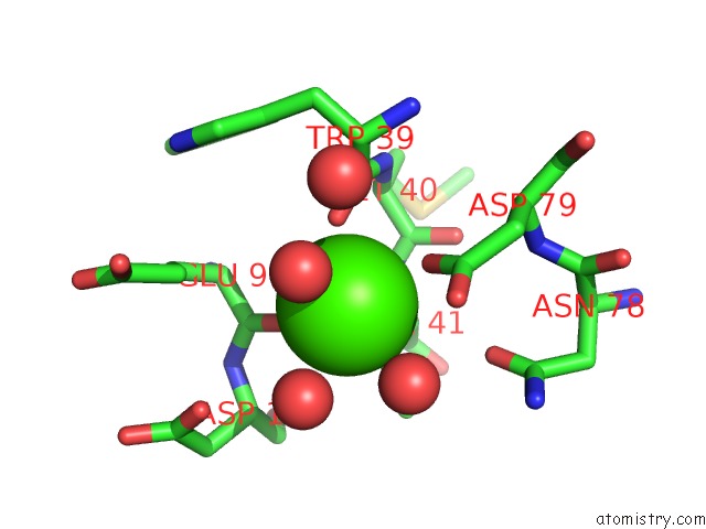
Mono view
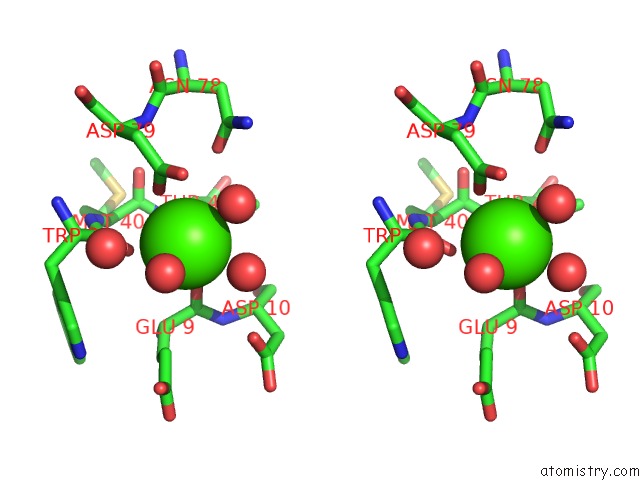
Stereo pair view
Mono view
Stereo pair view
A full contact list of Calcium with other atoms in the Ca binding
site number 1 of Crystal Structure of A Mutant T82R of A Betagamma-Crystallin Domain From Clostridium Beijerinckii within 5.0Å range:
|
Reference:
A.Mishra,
S.K.Suman,
S.S.Srivastava,
R.Sankaranarayanan,
Y.Sharma.
Decoding the Molecular Design Principles Underlying Ca(2+) Binding to Beta Gamma-Crystallin Motifs J.Mol.Biol. V. 415 75 2012.
ISSN: ISSN 0022-2836
PubMed: 22099475
DOI: 10.1016/J.JMB.2011.10.037
Page generated: Sat Jul 13 19:19:24 2024
ISSN: ISSN 0022-2836
PubMed: 22099475
DOI: 10.1016/J.JMB.2011.10.037
Last articles
Zn in 9J0NZn in 9J0O
Zn in 9J0P
Zn in 9FJX
Zn in 9EKB
Zn in 9C0F
Zn in 9CAH
Zn in 9CH0
Zn in 9CH3
Zn in 9CH1