Calcium »
PDB 3so0-3t2i »
3son »
Calcium in PDB 3son: Crystal Structure of A Putativel Nudix Hydrolase (LMOF2365_2679) From Listeria Monocytogenes Str. 4B F2365 at 1.70 A Resolution
Protein crystallography data
The structure of Crystal Structure of A Putativel Nudix Hydrolase (LMOF2365_2679) From Listeria Monocytogenes Str. 4B F2365 at 1.70 A Resolution, PDB code: 3son
was solved by
Joint Center For Structural Genomics (Jcsg),
with X-Ray Crystallography technique. A brief refinement statistics is given in the table below:
| Resolution Low / High (Å) | 27.67 / 1.71 |
| Space group | P 43 |
| Cell size a, b, c (Å), α, β, γ (°) | 57.880, 57.880, 94.490, 90.00, 90.00, 90.00 |
| R / Rfree (%) | 16.3 / 19.6 |
Calcium Binding Sites:
The binding sites of Calcium atom in the Crystal Structure of A Putativel Nudix Hydrolase (LMOF2365_2679) From Listeria Monocytogenes Str. 4B F2365 at 1.70 A Resolution
(pdb code 3son). This binding sites where shown within
5.0 Angstroms radius around Calcium atom.
In total 2 binding sites of Calcium where determined in the Crystal Structure of A Putativel Nudix Hydrolase (LMOF2365_2679) From Listeria Monocytogenes Str. 4B F2365 at 1.70 A Resolution, PDB code: 3son:
Jump to Calcium binding site number: 1; 2;
In total 2 binding sites of Calcium where determined in the Crystal Structure of A Putativel Nudix Hydrolase (LMOF2365_2679) From Listeria Monocytogenes Str. 4B F2365 at 1.70 A Resolution, PDB code: 3son:
Jump to Calcium binding site number: 1; 2;
Calcium binding site 1 out of 2 in 3son
Go back to
Calcium binding site 1 out
of 2 in the Crystal Structure of A Putativel Nudix Hydrolase (LMOF2365_2679) From Listeria Monocytogenes Str. 4B F2365 at 1.70 A Resolution
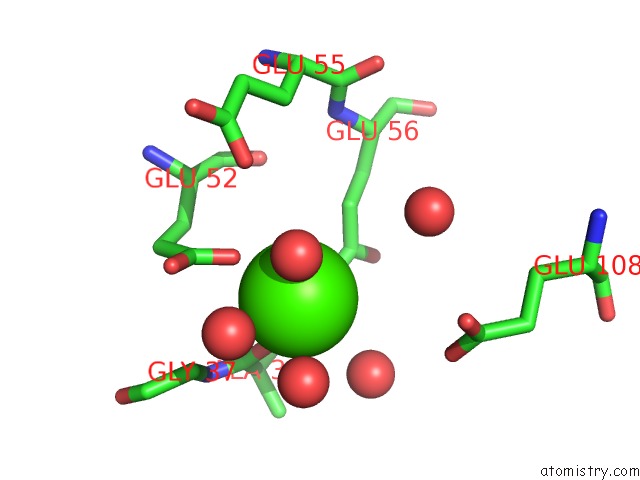
Mono view
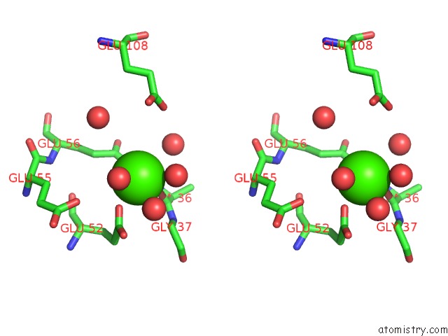
Stereo pair view
Mono view
Stereo pair view
A full contact list of Calcium with other atoms in the Ca binding
site number 1 of Crystal Structure of A Putativel Nudix Hydrolase (LMOF2365_2679) From Listeria Monocytogenes Str. 4B F2365 at 1.70 A Resolution within 5.0Å range:
|
Calcium binding site 2 out of 2 in 3son
Go back to
Calcium binding site 2 out
of 2 in the Crystal Structure of A Putativel Nudix Hydrolase (LMOF2365_2679) From Listeria Monocytogenes Str. 4B F2365 at 1.70 A Resolution
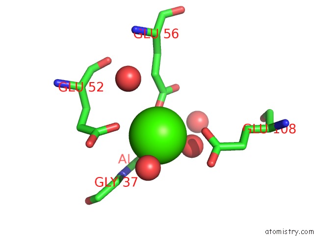
Mono view
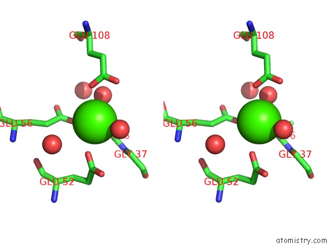
Stereo pair view
Mono view
Stereo pair view
A full contact list of Calcium with other atoms in the Ca binding
site number 2 of Crystal Structure of A Putativel Nudix Hydrolase (LMOF2365_2679) From Listeria Monocytogenes Str. 4B F2365 at 1.70 A Resolution within 5.0Å range:
|
Reference:
Joint Center For Structural Genomics (Jcsg),
Joint Center For Structural Genomics (Jcsg).
N/A N/A.
Page generated: Tue Jul 8 16:42:06 2025
Last articles
F in 7NTHF in 7NTI
F in 7NPC
F in 7NRG
F in 7NR5
F in 7NQS
F in 7NOS
F in 7NP5
F in 7NDV
F in 7NP6