Calcium »
PDB 3so0-3t2i »
3sqg »
Calcium in PDB 3sqg: Crystal Structure of A Methyl-Coenzyme M Reductase Purified From Black Sea Mats
Enzymatic activity of Crystal Structure of A Methyl-Coenzyme M Reductase Purified From Black Sea Mats
All present enzymatic activity of Crystal Structure of A Methyl-Coenzyme M Reductase Purified From Black Sea Mats:
2.8.4.1;
2.8.4.1;
Protein crystallography data
The structure of Crystal Structure of A Methyl-Coenzyme M Reductase Purified From Black Sea Mats, PDB code: 3sqg
was solved by
S.Shima,
M.Krueger,
T.Weinert,
U.Demmer,
R.K.Thauer,
U.Ermler,
with X-Ray Crystallography technique. A brief refinement statistics is given in the table below:
| Resolution Low / High (Å) | 47.58 / 2.10 |
| Space group | C 2 2 21 |
| Cell size a, b, c (Å), α, β, γ (°) | 128.860, 412.490, 165.510, 90.00, 90.00, 90.00 |
| R / Rfree (%) | 16.1 / 20.6 |
Other elements in 3sqg:
The structure of Crystal Structure of A Methyl-Coenzyme M Reductase Purified From Black Sea Mats also contains other interesting chemical elements:
| Nickel | (Ni) | 3 atoms |
| Chlorine | (Cl) | 1 atom |
Calcium Binding Sites:
The binding sites of Calcium atom in the Crystal Structure of A Methyl-Coenzyme M Reductase Purified From Black Sea Mats
(pdb code 3sqg). This binding sites where shown within
5.0 Angstroms radius around Calcium atom.
In total 2 binding sites of Calcium where determined in the Crystal Structure of A Methyl-Coenzyme M Reductase Purified From Black Sea Mats, PDB code: 3sqg:
Jump to Calcium binding site number: 1; 2;
In total 2 binding sites of Calcium where determined in the Crystal Structure of A Methyl-Coenzyme M Reductase Purified From Black Sea Mats, PDB code: 3sqg:
Jump to Calcium binding site number: 1; 2;
Calcium binding site 1 out of 2 in 3sqg
Go back to
Calcium binding site 1 out
of 2 in the Crystal Structure of A Methyl-Coenzyme M Reductase Purified From Black Sea Mats
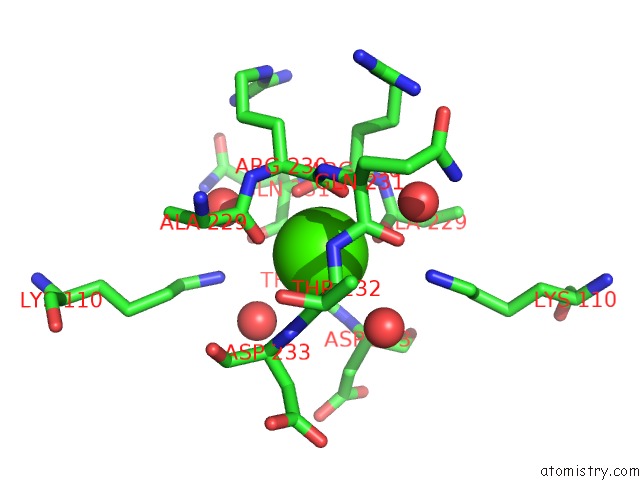
Mono view
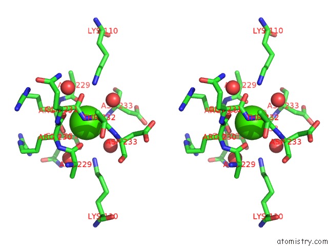
Stereo pair view
Mono view
Stereo pair view
A full contact list of Calcium with other atoms in the Ca binding
site number 1 of Crystal Structure of A Methyl-Coenzyme M Reductase Purified From Black Sea Mats within 5.0Å range:
|
Calcium binding site 2 out of 2 in 3sqg
Go back to
Calcium binding site 2 out
of 2 in the Crystal Structure of A Methyl-Coenzyme M Reductase Purified From Black Sea Mats
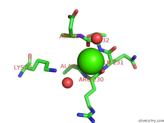
Mono view
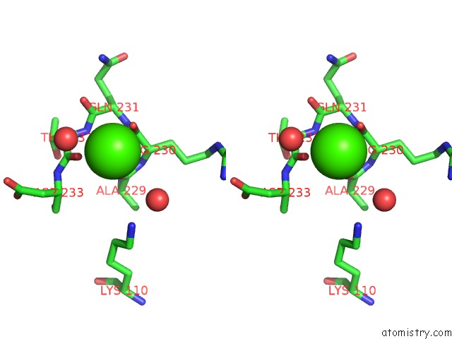
Stereo pair view
Mono view
Stereo pair view
A full contact list of Calcium with other atoms in the Ca binding
site number 2 of Crystal Structure of A Methyl-Coenzyme M Reductase Purified From Black Sea Mats within 5.0Å range:
|
Reference:
S.Shima,
M.Krueger,
T.Weinert,
U.Demmer,
J.Kahnt,
R.K.Thauer,
U.Ermler.
Structure of A Methyl-Coenzyme M Reductase From Black Sea Mats That Oxidize Methane Anaerobically. Nature V. 481 98 2011.
ISSN: ISSN 0028-0836
PubMed: 22121022
DOI: 10.1038/NATURE10663
Page generated: Tue Jul 8 16:44:24 2025
ISSN: ISSN 0028-0836
PubMed: 22121022
DOI: 10.1038/NATURE10663
Last articles
Ca in 7MG7Ca in 7MG6
Ca in 7MG5
Ca in 7MG4
Ca in 7MG3
Ca in 7MG2
Ca in 7MG1
Ca in 7MFL
Ca in 7MBS
Ca in 7MBV