Calcium »
PDB 3t2j-3ti3 »
3tey »
Calcium in PDB 3tey: Crystal Structure of Anthrax Protective Antigen (Membrane Insertion Loop Deleted) Mutant S337C N664C to 2.06-A Resolution
Protein crystallography data
The structure of Crystal Structure of Anthrax Protective Antigen (Membrane Insertion Loop Deleted) Mutant S337C N664C to 2.06-A Resolution, PDB code: 3tey
was solved by
G.K.Feld,
B.A.Krantz,
with X-Ray Crystallography technique. A brief refinement statistics is given in the table below:
| Resolution Low / High (Å) | 23.86 / 2.12 |
| Space group | P 21 21 21 |
| Cell size a, b, c (Å), α, β, γ (°) | 71.563, 94.602, 119.804, 90.00, 90.00, 90.00 |
| R / Rfree (%) | 20.7 / 23.4 |
Calcium Binding Sites:
The binding sites of Calcium atom in the Crystal Structure of Anthrax Protective Antigen (Membrane Insertion Loop Deleted) Mutant S337C N664C to 2.06-A Resolution
(pdb code 3tey). This binding sites where shown within
5.0 Angstroms radius around Calcium atom.
In total 2 binding sites of Calcium where determined in the Crystal Structure of Anthrax Protective Antigen (Membrane Insertion Loop Deleted) Mutant S337C N664C to 2.06-A Resolution, PDB code: 3tey:
Jump to Calcium binding site number: 1; 2;
In total 2 binding sites of Calcium where determined in the Crystal Structure of Anthrax Protective Antigen (Membrane Insertion Loop Deleted) Mutant S337C N664C to 2.06-A Resolution, PDB code: 3tey:
Jump to Calcium binding site number: 1; 2;
Calcium binding site 1 out of 2 in 3tey
Go back to
Calcium binding site 1 out
of 2 in the Crystal Structure of Anthrax Protective Antigen (Membrane Insertion Loop Deleted) Mutant S337C N664C to 2.06-A Resolution
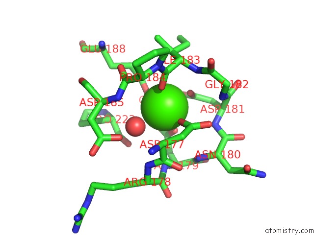
Mono view
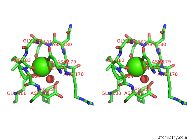
Stereo pair view
Mono view
Stereo pair view
A full contact list of Calcium with other atoms in the Ca binding
site number 1 of Crystal Structure of Anthrax Protective Antigen (Membrane Insertion Loop Deleted) Mutant S337C N664C to 2.06-A Resolution within 5.0Å range:
|
Calcium binding site 2 out of 2 in 3tey
Go back to
Calcium binding site 2 out
of 2 in the Crystal Structure of Anthrax Protective Antigen (Membrane Insertion Loop Deleted) Mutant S337C N664C to 2.06-A Resolution
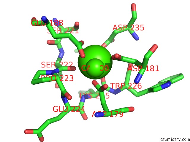
Mono view
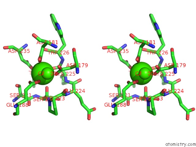
Stereo pair view
Mono view
Stereo pair view
A full contact list of Calcium with other atoms in the Ca binding
site number 2 of Crystal Structure of Anthrax Protective Antigen (Membrane Insertion Loop Deleted) Mutant S337C N664C to 2.06-A Resolution within 5.0Å range:
|
Reference:
G.K.Feld,
A.F.Kintzer,
I.I.Tang,
K.L.Thoren,
B.A.Krantz.
Domain Flexibility Modulates the Heterogeneous Assembly Mechanism of Anthrax Toxin Protective Antigen. J.Mol.Biol. V. 415 159 2012.
ISSN: ISSN 0022-2836
PubMed: 22063095
DOI: 10.1016/J.JMB.2011.10.035
Page generated: Tue Jul 8 16:55:51 2025
ISSN: ISSN 0022-2836
PubMed: 22063095
DOI: 10.1016/J.JMB.2011.10.035
Last articles
Ca in 4DTUCa in 4DTX
Ca in 4DTS
Ca in 4DTR
Ca in 4DTN
Ca in 4DTO
Ca in 4DTP
Ca in 4DTM
Ca in 4DTJ
Ca in 4DSK