Calcium »
PDB 3ti4-3tvc »
3tic »
Calcium in PDB 3tic: Crystal Structure of 1957 Pandemic H2N2 Neuraminidase Complexed with Zanamivir
Protein crystallography data
The structure of Crystal Structure of 1957 Pandemic H2N2 Neuraminidase Complexed with Zanamivir, PDB code: 3tic
was solved by
C.J.Vavricka,
Q.Li,
Y.Wu,
J.Qi,
M.Wang,
Y.Liu,
F.Gao,
J.Liu,
E.Feng,
J.He,
J.Wang,
H.Liu,
H.Jiang,
G.F.Gao,
with X-Ray Crystallography technique. A brief refinement statistics is given in the table below:
| Resolution Low / High (Å) | 41.27 / 1.89 |
| Space group | P 1 21 1 |
| Cell size a, b, c (Å), α, β, γ (°) | 90.129, 139.994, 90.173, 90.00, 101.34, 90.00 |
| R / Rfree (%) | 15.3 / 18.4 |
Calcium Binding Sites:
The binding sites of Calcium atom in the Crystal Structure of 1957 Pandemic H2N2 Neuraminidase Complexed with Zanamivir
(pdb code 3tic). This binding sites where shown within
5.0 Angstroms radius around Calcium atom.
In total 4 binding sites of Calcium where determined in the Crystal Structure of 1957 Pandemic H2N2 Neuraminidase Complexed with Zanamivir, PDB code: 3tic:
Jump to Calcium binding site number: 1; 2; 3; 4;
In total 4 binding sites of Calcium where determined in the Crystal Structure of 1957 Pandemic H2N2 Neuraminidase Complexed with Zanamivir, PDB code: 3tic:
Jump to Calcium binding site number: 1; 2; 3; 4;
Calcium binding site 1 out of 4 in 3tic
Go back to
Calcium binding site 1 out
of 4 in the Crystal Structure of 1957 Pandemic H2N2 Neuraminidase Complexed with Zanamivir
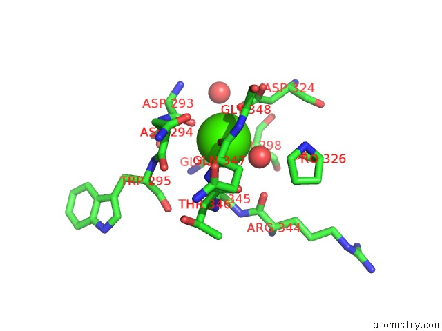
Mono view
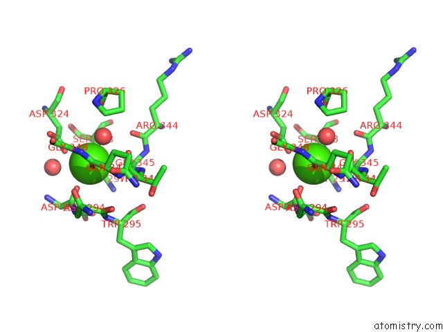
Stereo pair view
Mono view
Stereo pair view
A full contact list of Calcium with other atoms in the Ca binding
site number 1 of Crystal Structure of 1957 Pandemic H2N2 Neuraminidase Complexed with Zanamivir within 5.0Å range:
|
Calcium binding site 2 out of 4 in 3tic
Go back to
Calcium binding site 2 out
of 4 in the Crystal Structure of 1957 Pandemic H2N2 Neuraminidase Complexed with Zanamivir
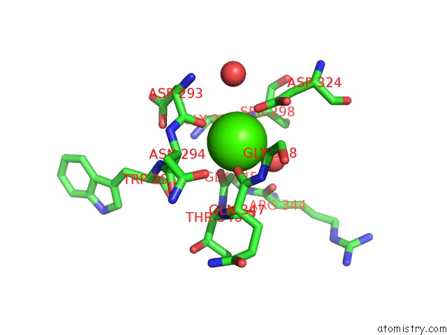
Mono view
Stereo pair view
Mono view
Stereo pair view
A full contact list of Calcium with other atoms in the Ca binding
site number 2 of Crystal Structure of 1957 Pandemic H2N2 Neuraminidase Complexed with Zanamivir within 5.0Å range:
|
Calcium binding site 3 out of 4 in 3tic
Go back to
Calcium binding site 3 out
of 4 in the Crystal Structure of 1957 Pandemic H2N2 Neuraminidase Complexed with Zanamivir

Mono view

Stereo pair view
Mono view
Stereo pair view
A full contact list of Calcium with other atoms in the Ca binding
site number 3 of Crystal Structure of 1957 Pandemic H2N2 Neuraminidase Complexed with Zanamivir within 5.0Å range:
|
Calcium binding site 4 out of 4 in 3tic
Go back to
Calcium binding site 4 out
of 4 in the Crystal Structure of 1957 Pandemic H2N2 Neuraminidase Complexed with Zanamivir

Mono view

Stereo pair view
Mono view
Stereo pair view
A full contact list of Calcium with other atoms in the Ca binding
site number 4 of Crystal Structure of 1957 Pandemic H2N2 Neuraminidase Complexed with Zanamivir within 5.0Å range:
|
Reference:
C.J.Vavricka,
Q.Li,
Y.Wu,
J.Qi,
M.Wang,
Y.Liu,
F.Gao,
J.Liu,
E.Feng,
J.He,
J.Wang,
H.Liu,
H.Jiang,
G.F.Gao.
Structural and Functional Analysis of Laninamivir and Its Octanoate Prodrug Reveals Group Specific Mechanisms For Influenza Na Inhibition Plos Pathog. V. 7 02249 2011.
ISSN: ISSN 1553-7366
PubMed: 22028647
DOI: 10.1371/JOURNAL.PPAT.1002249
Page generated: Sat Jul 13 19:47:07 2024
ISSN: ISSN 1553-7366
PubMed: 22028647
DOI: 10.1371/JOURNAL.PPAT.1002249
Last articles
Zn in 9J0NZn in 9J0O
Zn in 9J0P
Zn in 9FJX
Zn in 9EKB
Zn in 9C0F
Zn in 9CAH
Zn in 9CH0
Zn in 9CH3
Zn in 9CH1