Calcium »
PDB 3ti4-3tvc »
3tmn »
Calcium in PDB 3tmn: The Binding of L-Valyl-L-Tryptophan to Crystalline Thermolysin Illustrates the Mode of Interaction of A Product of Peptide Hydrolysis
Enzymatic activity of The Binding of L-Valyl-L-Tryptophan to Crystalline Thermolysin Illustrates the Mode of Interaction of A Product of Peptide Hydrolysis
All present enzymatic activity of The Binding of L-Valyl-L-Tryptophan to Crystalline Thermolysin Illustrates the Mode of Interaction of A Product of Peptide Hydrolysis:
3.4.24.27;
3.4.24.27;
Protein crystallography data
The structure of The Binding of L-Valyl-L-Tryptophan to Crystalline Thermolysin Illustrates the Mode of Interaction of A Product of Peptide Hydrolysis, PDB code: 3tmn
was solved by
H.M.Holden,
B.W.Matthews,
with X-Ray Crystallography technique. A brief refinement statistics is given in the table below:
| Resolution Low / High (Å) | 10.00 / 1.70 |
| Space group | P 61 2 2 |
| Cell size a, b, c (Å), α, β, γ (°) | 94.100, 94.100, 131.400, 90.00, 90.00, 120.00 |
| R / Rfree (%) | n/a / n/a |
Other elements in 3tmn:
The structure of The Binding of L-Valyl-L-Tryptophan to Crystalline Thermolysin Illustrates the Mode of Interaction of A Product of Peptide Hydrolysis also contains other interesting chemical elements:
| Zinc | (Zn) | 1 atom |
Calcium Binding Sites:
The binding sites of Calcium atom in the The Binding of L-Valyl-L-Tryptophan to Crystalline Thermolysin Illustrates the Mode of Interaction of A Product of Peptide Hydrolysis
(pdb code 3tmn). This binding sites where shown within
5.0 Angstroms radius around Calcium atom.
In total 4 binding sites of Calcium where determined in the The Binding of L-Valyl-L-Tryptophan to Crystalline Thermolysin Illustrates the Mode of Interaction of A Product of Peptide Hydrolysis, PDB code: 3tmn:
Jump to Calcium binding site number: 1; 2; 3; 4;
In total 4 binding sites of Calcium where determined in the The Binding of L-Valyl-L-Tryptophan to Crystalline Thermolysin Illustrates the Mode of Interaction of A Product of Peptide Hydrolysis, PDB code: 3tmn:
Jump to Calcium binding site number: 1; 2; 3; 4;
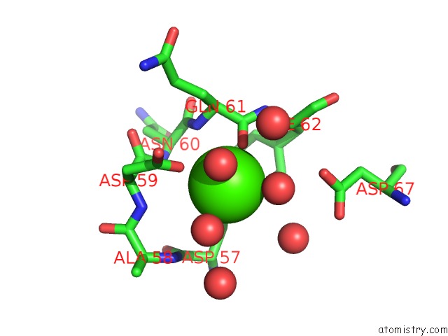
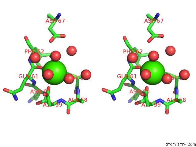
Calcium binding site 1 out of 4 in 3tmn
Go back to
Calcium binding site 1 out
of 4 in the The Binding of L-Valyl-L-Tryptophan to Crystalline Thermolysin Illustrates the Mode of Interaction of A Product of Peptide Hydrolysis
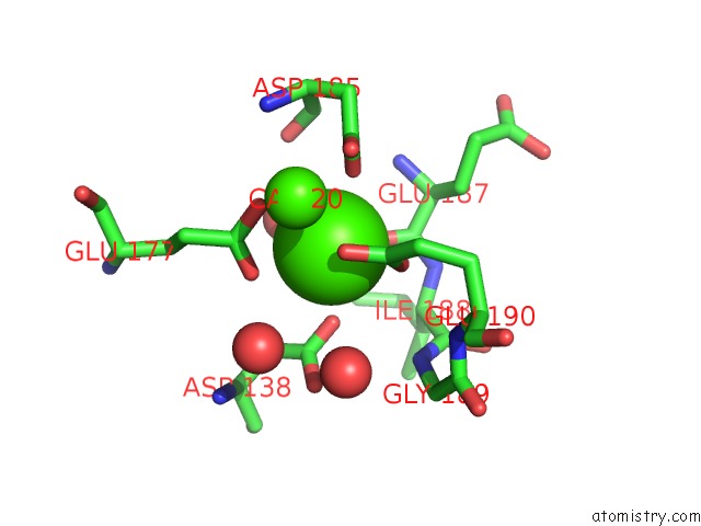
Mono view
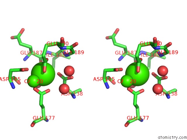
Stereo pair view
Mono view
Stereo pair view
A full contact list of Calcium with other atoms in the Ca binding
site number 1 of The Binding of L-Valyl-L-Tryptophan to Crystalline Thermolysin Illustrates the Mode of Interaction of A Product of Peptide Hydrolysis within 5.0Å range:
|
Calcium binding site 2 out of 4 in 3tmn
Go back to
Calcium binding site 2 out
of 4 in the The Binding of L-Valyl-L-Tryptophan to Crystalline Thermolysin Illustrates the Mode of Interaction of A Product of Peptide Hydrolysis
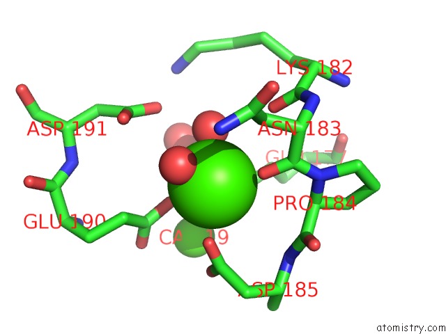
Mono view
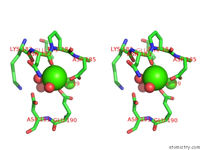
Stereo pair view
Mono view
Stereo pair view
A full contact list of Calcium with other atoms in the Ca binding
site number 2 of The Binding of L-Valyl-L-Tryptophan to Crystalline Thermolysin Illustrates the Mode of Interaction of A Product of Peptide Hydrolysis within 5.0Å range:
|
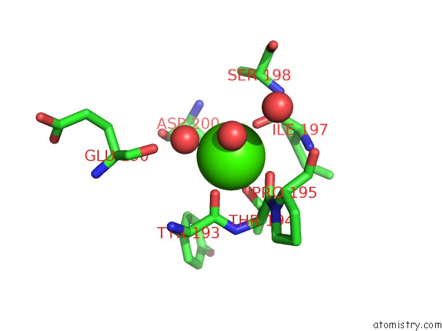
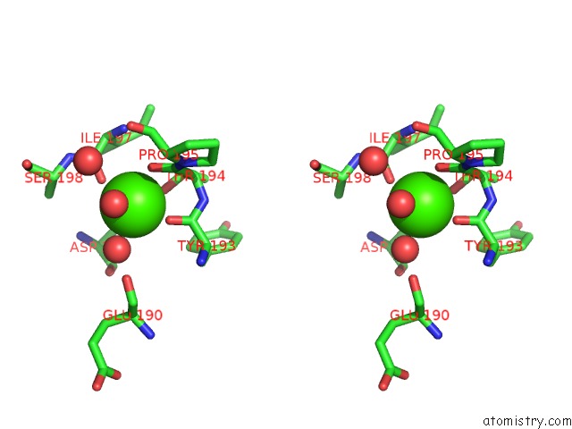
Calcium binding site 3 out of 4 in 3tmn
Go back to
Calcium binding site 3 out
of 4 in the The Binding of L-Valyl-L-Tryptophan to Crystalline Thermolysin Illustrates the Mode of Interaction of A Product of Peptide Hydrolysis
Mono view
Stereo pair view
Mono view
Stereo pair view
A full contact list of Calcium with other atoms in the Ca binding
site number 3 of The Binding of L-Valyl-L-Tryptophan to Crystalline Thermolysin Illustrates the Mode of Interaction of A Product of Peptide Hydrolysis within 5.0Å range:
|
Calcium binding site 4 out of 4 in 3tmn
Go back to
Calcium binding site 4 out
of 4 in the The Binding of L-Valyl-L-Tryptophan to Crystalline Thermolysin Illustrates the Mode of Interaction of A Product of Peptide Hydrolysis
Mono view
Stereo pair view
Mono view
Stereo pair view
A full contact list of Calcium with other atoms in the Ca binding
site number 4 of The Binding of L-Valyl-L-Tryptophan to Crystalline Thermolysin Illustrates the Mode of Interaction of A Product of Peptide Hydrolysis within 5.0Å range:
|
Reference:
H.M.Holden,
B.W.Matthews.
The Binding of L-Valyl-L-Tryptophan to Crystalline Thermolysin Illustrates the Mode of Interaction of A Product of Peptide Hydrolysis. J.Biol.Chem. V. 263 3256 1988.
ISSN: ISSN 0021-9258
PubMed: 3343246
Page generated: Tue Jul 8 17:01:42 2025
ISSN: ISSN 0021-9258
PubMed: 3343246
Last articles
Cl in 5LA5Cl in 5L9J
Cl in 5L9Z
Cl in 5L7I
Cl in 5L8D
Cl in 5L7U
Cl in 5L7P
Cl in 5L6I
Cl in 5L6H
Cl in 5L7F