Calcium »
PDB 3ti4-3tvc »
3tuy »
Calcium in PDB 3tuy: Phosphorylated Light Chain Domain of Scallop Smooth Muscle Myosin
Protein crystallography data
The structure of Phosphorylated Light Chain Domain of Scallop Smooth Muscle Myosin, PDB code: 3tuy
was solved by
V.S.S.Kumar,
E.O'neall-Hennessey,
L.Reshetnikova,
J.H.Brown,
H.Robinson,
A.G.Szent-Gyorgyi,
C.Cohen,
with X-Ray Crystallography technique. A brief refinement statistics is given in the table below:
| Resolution Low / High (Å) | 48.83 / 2.50 |
| Space group | P 1 |
| Cell size a, b, c (Å), α, β, γ (°) | 50.946, 69.232, 79.503, 77.20, 85.99, 73.44 |
| R / Rfree (%) | 20 / 25.2 |
Other elements in 3tuy:
The structure of Phosphorylated Light Chain Domain of Scallop Smooth Muscle Myosin also contains other interesting chemical elements:
| Magnesium | (Mg) | 2 atoms |
Calcium Binding Sites:
The binding sites of Calcium atom in the Phosphorylated Light Chain Domain of Scallop Smooth Muscle Myosin
(pdb code 3tuy). This binding sites where shown within
5.0 Angstroms radius around Calcium atom.
In total 2 binding sites of Calcium where determined in the Phosphorylated Light Chain Domain of Scallop Smooth Muscle Myosin, PDB code: 3tuy:
Jump to Calcium binding site number: 1; 2;
In total 2 binding sites of Calcium where determined in the Phosphorylated Light Chain Domain of Scallop Smooth Muscle Myosin, PDB code: 3tuy:
Jump to Calcium binding site number: 1; 2;
Calcium binding site 1 out of 2 in 3tuy
Go back to
Calcium binding site 1 out
of 2 in the Phosphorylated Light Chain Domain of Scallop Smooth Muscle Myosin
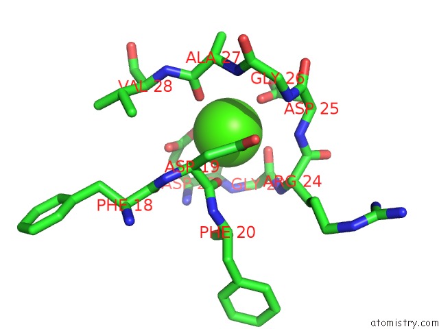
Mono view
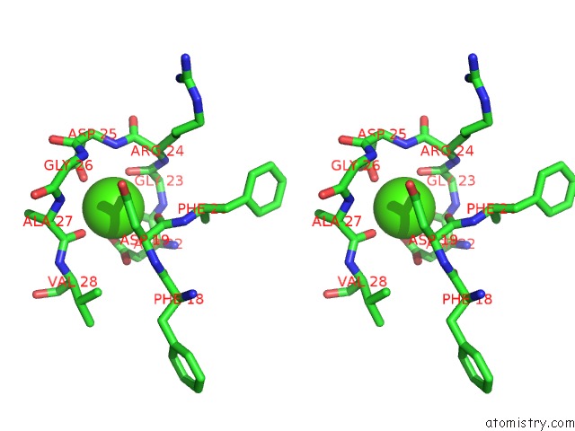
Stereo pair view
Mono view
Stereo pair view
A full contact list of Calcium with other atoms in the Ca binding
site number 1 of Phosphorylated Light Chain Domain of Scallop Smooth Muscle Myosin within 5.0Å range:
|
Calcium binding site 2 out of 2 in 3tuy
Go back to
Calcium binding site 2 out
of 2 in the Phosphorylated Light Chain Domain of Scallop Smooth Muscle Myosin
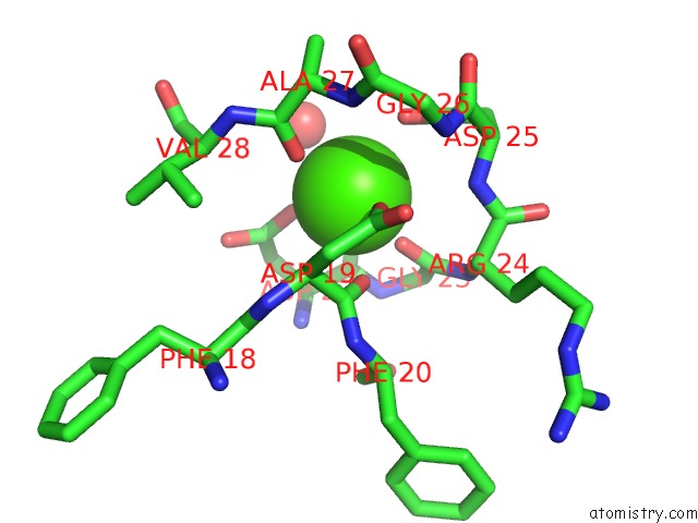
Mono view
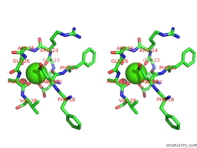
Stereo pair view
Mono view
Stereo pair view
A full contact list of Calcium with other atoms in the Ca binding
site number 2 of Phosphorylated Light Chain Domain of Scallop Smooth Muscle Myosin within 5.0Å range:
|
Reference:
V.S.Senthil Kumar,
E.O'neall-Hennessey,
L.Reshetnikova,
J.H.Brown,
H.Robinson,
A.G.Szent-Gyorgyi,
C.Cohen.
Crystal Structure of A Phosphorylated Light Chain Domain of Scallop Smooth-Muscle Myosin. Biophys.J. V. 101 2185 2011.
ISSN: ISSN 0006-3495
PubMed: 22067157
DOI: 10.1016/J.BPJ.2011.09.028
Page generated: Sat Jul 13 19:55:59 2024
ISSN: ISSN 0006-3495
PubMed: 22067157
DOI: 10.1016/J.BPJ.2011.09.028
Last articles
Zn in 9JYWZn in 9IR4
Zn in 9IR3
Zn in 9GMX
Zn in 9GMW
Zn in 9JEJ
Zn in 9ERF
Zn in 9ERE
Zn in 9EGV
Zn in 9EGW