Calcium »
PDB 3tz1-3uiq »
3u39 »
Calcium in PDB 3u39: Crystal Structure of the Apo Bacillus Stearothermophilus Phosphofructokinase
Enzymatic activity of Crystal Structure of the Apo Bacillus Stearothermophilus Phosphofructokinase
All present enzymatic activity of Crystal Structure of the Apo Bacillus Stearothermophilus Phosphofructokinase:
2.7.1.11;
2.7.1.11;
Protein crystallography data
The structure of Crystal Structure of the Apo Bacillus Stearothermophilus Phosphofructokinase, PDB code: 3u39
was solved by
R.Mosser,
M.C.M.Reddy,
J.B.Bruning,
J.C.Sacchettini,
G.D.Reinhart,
with X-Ray Crystallography technique. A brief refinement statistics is given in the table below:
| Resolution Low / High (Å) | 28.51 / 2.79 |
| Space group | C 1 2 1 |
| Cell size a, b, c (Å), α, β, γ (°) | 201.909, 113.383, 76.627, 90.00, 104.03, 90.00 |
| R / Rfree (%) | 19.9 / 26 |
Calcium Binding Sites:
The binding sites of Calcium atom in the Crystal Structure of the Apo Bacillus Stearothermophilus Phosphofructokinase
(pdb code 3u39). This binding sites where shown within
5.0 Angstroms radius around Calcium atom.
In total 5 binding sites of Calcium where determined in the Crystal Structure of the Apo Bacillus Stearothermophilus Phosphofructokinase, PDB code: 3u39:
Jump to Calcium binding site number: 1; 2; 3; 4; 5;
In total 5 binding sites of Calcium where determined in the Crystal Structure of the Apo Bacillus Stearothermophilus Phosphofructokinase, PDB code: 3u39:
Jump to Calcium binding site number: 1; 2; 3; 4; 5;
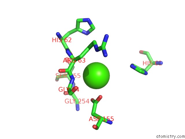
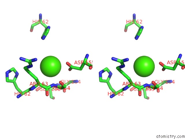
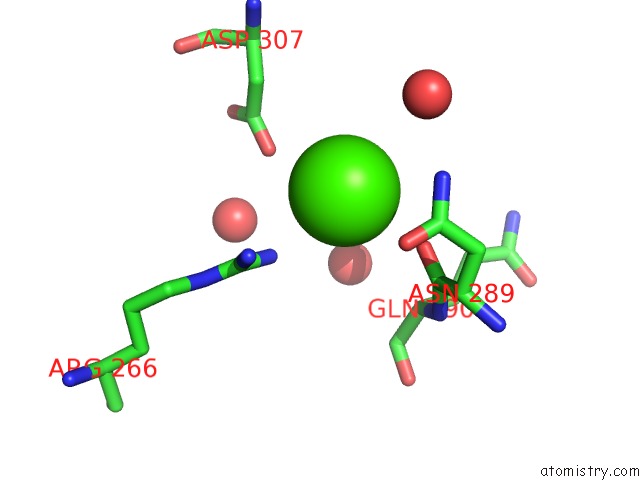
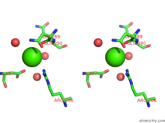
Calcium binding site 1 out of 5 in 3u39
Go back to
Calcium binding site 1 out
of 5 in the Crystal Structure of the Apo Bacillus Stearothermophilus Phosphofructokinase
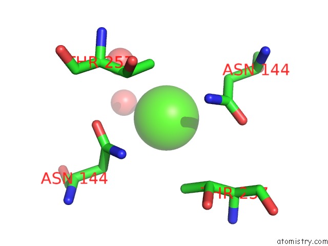
Mono view
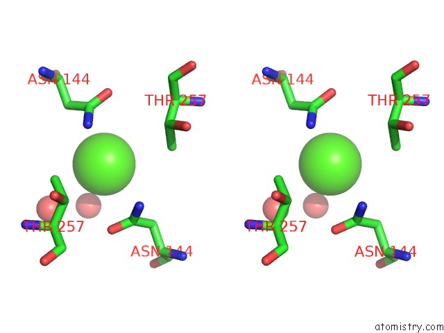
Stereo pair view
Mono view
Stereo pair view
A full contact list of Calcium with other atoms in the Ca binding
site number 1 of Crystal Structure of the Apo Bacillus Stearothermophilus Phosphofructokinase within 5.0Å range:
|
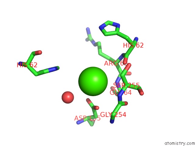
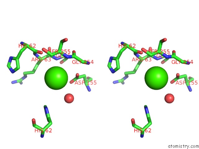
Calcium binding site 2 out of 5 in 3u39
Go back to
Calcium binding site 2 out
of 5 in the Crystal Structure of the Apo Bacillus Stearothermophilus Phosphofructokinase
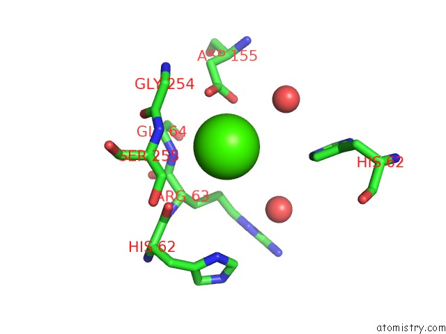
Mono view
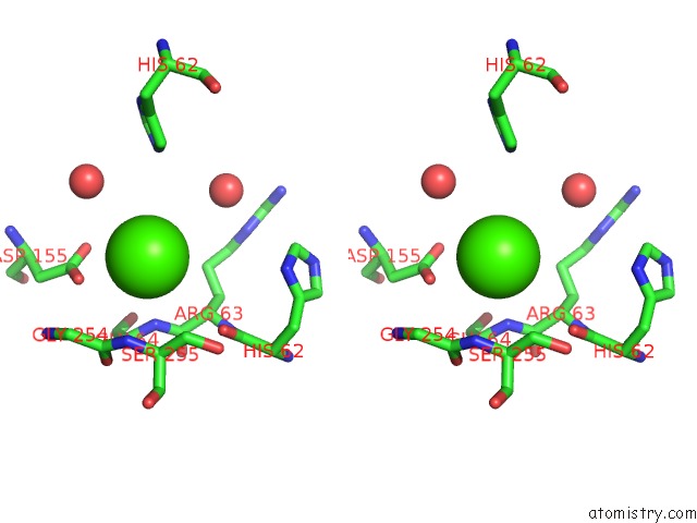
Stereo pair view
Mono view
Stereo pair view
A full contact list of Calcium with other atoms in the Ca binding
site number 2 of Crystal Structure of the Apo Bacillus Stearothermophilus Phosphofructokinase within 5.0Å range:
|
Calcium binding site 3 out of 5 in 3u39
Go back to
Calcium binding site 3 out
of 5 in the Crystal Structure of the Apo Bacillus Stearothermophilus Phosphofructokinase
Mono view
Stereo pair view
Mono view
Stereo pair view
A full contact list of Calcium with other atoms in the Ca binding
site number 3 of Crystal Structure of the Apo Bacillus Stearothermophilus Phosphofructokinase within 5.0Å range:
|
Calcium binding site 4 out of 5 in 3u39
Go back to
Calcium binding site 4 out
of 5 in the Crystal Structure of the Apo Bacillus Stearothermophilus Phosphofructokinase
Mono view
Stereo pair view
Mono view
Stereo pair view
A full contact list of Calcium with other atoms in the Ca binding
site number 4 of Crystal Structure of the Apo Bacillus Stearothermophilus Phosphofructokinase within 5.0Å range:
|
Calcium binding site 5 out of 5 in 3u39
Go back to
Calcium binding site 5 out
of 5 in the Crystal Structure of the Apo Bacillus Stearothermophilus Phosphofructokinase
Mono view
Stereo pair view
Mono view
Stereo pair view
A full contact list of Calcium with other atoms in the Ca binding
site number 5 of Crystal Structure of the Apo Bacillus Stearothermophilus Phosphofructokinase within 5.0Å range:
|
Reference:
R.Mosser,
M.C.Reddy,
J.B.Bruning,
J.C.Sacchettini,
G.D.Reinhart.
Structure of the Apo Form of Bacillus Stearothermophilus Phosphofructokinase. Biochemistry V. 51 769 2012.
ISSN: ISSN 0006-2960
PubMed: 22212099
DOI: 10.1021/BI201548P
Page generated: Sat Jul 13 19:58:48 2024
ISSN: ISSN 0006-2960
PubMed: 22212099
DOI: 10.1021/BI201548P
Last articles
Zn in 9MJ5Zn in 9HNW
Zn in 9G0L
Zn in 9FNE
Zn in 9DZN
Zn in 9E0I
Zn in 9D32
Zn in 9DAK
Zn in 8ZXC
Zn in 8ZUF