Calcium »
PDB 3tz1-3uiq »
3ub5 »
Calcium in PDB 3ub5: Profilin:Actin with A Wide Open Nucleotide Cleft
Protein crystallography data
The structure of Profilin:Actin with A Wide Open Nucleotide Cleft, PDB code: 3ub5
was solved by
J.C.Porta,
G.E.Borgstahl,
with X-Ray Crystallography technique. A brief refinement statistics is given in the table below:
| Resolution Low / High (Å) | 37.24 / 2.20 |
| Space group | P 21 21 21 |
| Cell size a, b, c (Å), α, β, γ (°) | 38.000, 72.000, 186.800, 90.00, 90.00, 90.00 |
| R / Rfree (%) | 21.5 / 29 |
Other elements in 3ub5:
The structure of Profilin:Actin with A Wide Open Nucleotide Cleft also contains other interesting chemical elements:
| Chlorine | (Cl) | 1 atom |
Calcium Binding Sites:
The binding sites of Calcium atom in the Profilin:Actin with A Wide Open Nucleotide Cleft
(pdb code 3ub5). This binding sites where shown within
5.0 Angstroms radius around Calcium atom.
In total 2 binding sites of Calcium where determined in the Profilin:Actin with A Wide Open Nucleotide Cleft, PDB code: 3ub5:
Jump to Calcium binding site number: 1; 2;
In total 2 binding sites of Calcium where determined in the Profilin:Actin with A Wide Open Nucleotide Cleft, PDB code: 3ub5:
Jump to Calcium binding site number: 1; 2;
Calcium binding site 1 out of 2 in 3ub5
Go back to
Calcium binding site 1 out
of 2 in the Profilin:Actin with A Wide Open Nucleotide Cleft
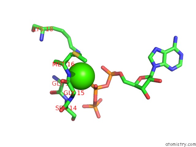
Mono view
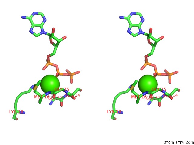
Stereo pair view
Mono view
Stereo pair view
A full contact list of Calcium with other atoms in the Ca binding
site number 1 of Profilin:Actin with A Wide Open Nucleotide Cleft within 5.0Å range:
|
Calcium binding site 2 out of 2 in 3ub5
Go back to
Calcium binding site 2 out
of 2 in the Profilin:Actin with A Wide Open Nucleotide Cleft
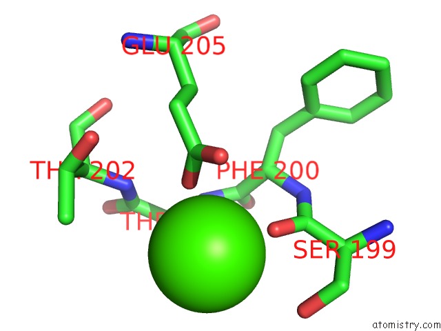
Mono view
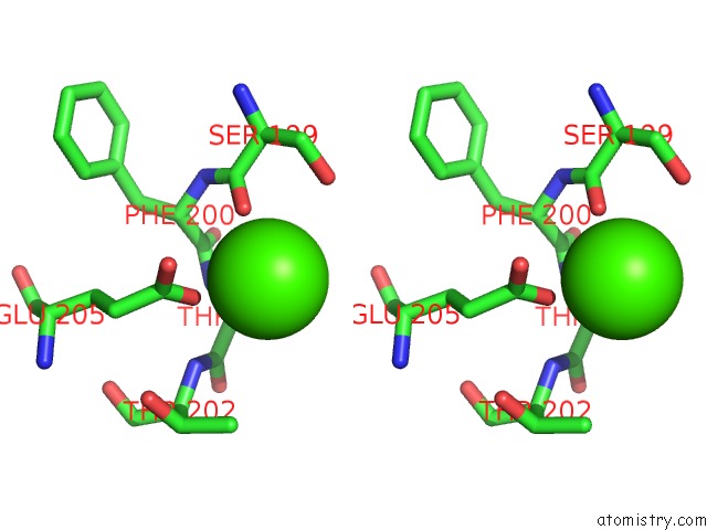
Stereo pair view
Mono view
Stereo pair view
A full contact list of Calcium with other atoms in the Ca binding
site number 2 of Profilin:Actin with A Wide Open Nucleotide Cleft within 5.0Å range:
|
Reference:
J.C.Porta,
G.E.Borgstahl.
Structural Basis For Profilin-Mediated Actin Nucleotide Exchange. J.Mol.Biol. V. 418 103 2012.
ISSN: ISSN 0022-2836
PubMed: 22366544
DOI: 10.1016/J.JMB.2012.02.012
Page generated: Tue Jul 8 17:12:45 2025
ISSN: ISSN 0022-2836
PubMed: 22366544
DOI: 10.1016/J.JMB.2012.02.012
Last articles
Cl in 5R9CCl in 5R9E
Cl in 5R9B
Cl in 5R9D
Cl in 5R9A
Cl in 5R99
Cl in 5R98
Cl in 5R96
Cl in 5R97
Cl in 5R95