Calcium »
PDB 3tz1-3uiq »
3ubf »
Calcium in PDB 3ubf: Crystal Structure of Drosophila N-Cadherin EC1-3, I
Protein crystallography data
The structure of Crystal Structure of Drosophila N-Cadherin EC1-3, I, PDB code: 3ubf
was solved by
X.Jin,
M.A.Walker,
L.Shapiro,
with X-Ray Crystallography technique. A brief refinement statistics is given in the table below:
| Resolution Low / High (Å) | 20.00 / 2.50 |
| Space group | P 62 2 2 |
| Cell size a, b, c (Å), α, β, γ (°) | 96.289, 96.289, 148.196, 90.00, 90.00, 120.00 |
| R / Rfree (%) | 20.1 / 27.9 |
Other elements in 3ubf:
The structure of Crystal Structure of Drosophila N-Cadherin EC1-3, I also contains other interesting chemical elements:
| Zinc | (Zn) | 18 atoms |
Calcium Binding Sites:
The binding sites of Calcium atom in the Crystal Structure of Drosophila N-Cadherin EC1-3, I
(pdb code 3ubf). This binding sites where shown within
5.0 Angstroms radius around Calcium atom.
In total only one binding site of Calcium was determined in the Crystal Structure of Drosophila N-Cadherin EC1-3, I, PDB code: 3ubf:
In total only one binding site of Calcium was determined in the Crystal Structure of Drosophila N-Cadherin EC1-3, I, PDB code: 3ubf:
Calcium binding site 1 out of 1 in 3ubf
Go back to
Calcium binding site 1 out
of 1 in the Crystal Structure of Drosophila N-Cadherin EC1-3, I
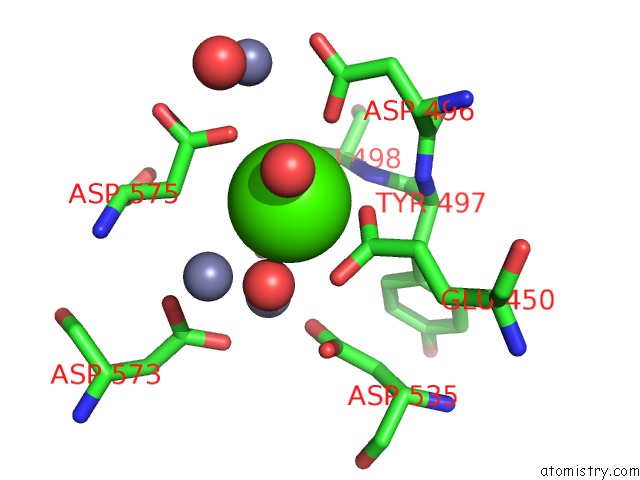
Mono view
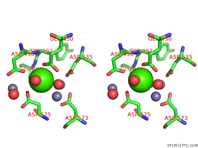
Stereo pair view
Mono view
Stereo pair view
A full contact list of Calcium with other atoms in the Ca binding
site number 1 of Crystal Structure of Drosophila N-Cadherin EC1-3, I within 5.0Å range:
|
Reference:
X.Jin,
M.A.Walker,
K.Felsovalyi,
J.Vendome,
F.Bahna,
S.Mannepalli,
F.Cosmanescu,
G.Ahlsen,
B.Honig,
L.Shapiro.
Crystal Structures of Drosophila N-Cadherin Ectodomain Regions Reveal A Widely Used Class of CA2+-Free Interdomain Linkers. Proc.Natl.Acad.Sci.Usa V. 109 E127 2012.
ISSN: ISSN 0027-8424
PubMed: 22171007
DOI: 10.1073/PNAS.1117538108
Page generated: Tue Jul 8 17:13:15 2025
ISSN: ISSN 0027-8424
PubMed: 22171007
DOI: 10.1073/PNAS.1117538108
Last articles
Fe in 2YXOFe in 2YRS
Fe in 2YXC
Fe in 2YNM
Fe in 2YVJ
Fe in 2YP1
Fe in 2YU2
Fe in 2YU1
Fe in 2YQB
Fe in 2YOO