Calcium »
PDB 3uix-3v03 »
3uqo »
Calcium in PDB 3uqo: Bovine Trypsin Variant X(TRIPLEPHE227) in Complex with Small Molecule Inhibitor
Enzymatic activity of Bovine Trypsin Variant X(TRIPLEPHE227) in Complex with Small Molecule Inhibitor
All present enzymatic activity of Bovine Trypsin Variant X(TRIPLEPHE227) in Complex with Small Molecule Inhibitor:
3.4.21.4;
3.4.21.4;
Protein crystallography data
The structure of Bovine Trypsin Variant X(TRIPLEPHE227) in Complex with Small Molecule Inhibitor, PDB code: 3uqo
was solved by
A.Tziridis,
P.Neumann,
P.Kolenko,
M.T.Stubbs,
with X-Ray Crystallography technique. A brief refinement statistics is given in the table below:
| Resolution Low / High (Å) | 30.00 / 1.80 |
| Space group | P 32 2 1 |
| Cell size a, b, c (Å), α, β, γ (°) | 54.692, 54.692, 112.953, 90.00, 90.00, 120.00 |
| R / Rfree (%) | 20 / 25.6 |
Calcium Binding Sites:
The binding sites of Calcium atom in the Bovine Trypsin Variant X(TRIPLEPHE227) in Complex with Small Molecule Inhibitor
(pdb code 3uqo). This binding sites where shown within
5.0 Angstroms radius around Calcium atom.
In total only one binding site of Calcium was determined in the Bovine Trypsin Variant X(TRIPLEPHE227) in Complex with Small Molecule Inhibitor, PDB code: 3uqo:
In total only one binding site of Calcium was determined in the Bovine Trypsin Variant X(TRIPLEPHE227) in Complex with Small Molecule Inhibitor, PDB code: 3uqo:
Calcium binding site 1 out of 1 in 3uqo
Go back to
Calcium binding site 1 out
of 1 in the Bovine Trypsin Variant X(TRIPLEPHE227) in Complex with Small Molecule Inhibitor
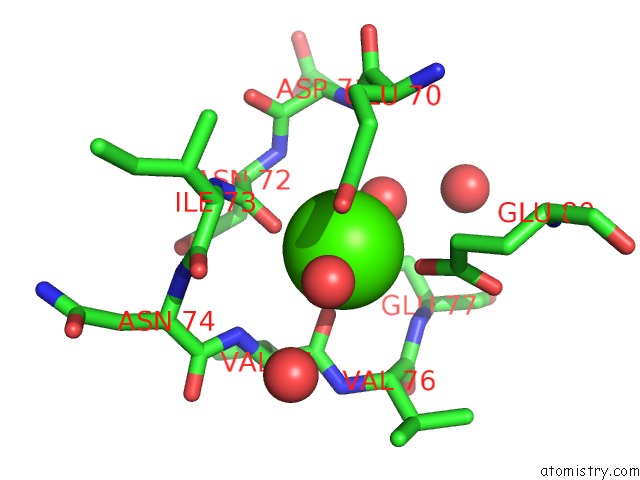
Mono view
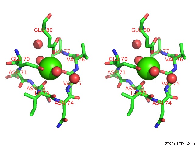
Stereo pair view
Mono view
Stereo pair view
A full contact list of Calcium with other atoms in the Ca binding
site number 1 of Bovine Trypsin Variant X(TRIPLEPHE227) in Complex with Small Molecule Inhibitor within 5.0Å range:
|
Reference:
A.Tziridis,
D.Rauh,
P.Neumann,
P.Kolenko,
A.Menzel,
U.Brauer,
C.Ursel,
P.Steinmetzer,
J.Sturzebecher,
A.Schweinitz,
T.Steinmetzer,
M.T.Stubbs.
Correlating Structure and Ligand Affinity in Drug Discovery: A Cautionary Tale Involving Second Shell Residues. Biol.Chem. V. 395 891 2014.
ISSN: ISSN 1431-6730
PubMed: 25003390
DOI: 10.1515/HSZ-2014-0158
Page generated: Tue Jul 8 17:19:03 2025
ISSN: ISSN 1431-6730
PubMed: 25003390
DOI: 10.1515/HSZ-2014-0158
Last articles
Fe in 2YXOFe in 2YRS
Fe in 2YXC
Fe in 2YNM
Fe in 2YVJ
Fe in 2YP1
Fe in 2YU2
Fe in 2YU1
Fe in 2YQB
Fe in 2YOO