Calcium »
PDB 3v0a-3vl2 »
3vdr »
Calcium in PDB 3vdr: Crystal Structure of D-3-Hydroxybutyrate Dehydrogenase, Prepared in the Presence of the Substrate D-3-Hydroxybutyrate and Nad(+)
Enzymatic activity of Crystal Structure of D-3-Hydroxybutyrate Dehydrogenase, Prepared in the Presence of the Substrate D-3-Hydroxybutyrate and Nad(+)
All present enzymatic activity of Crystal Structure of D-3-Hydroxybutyrate Dehydrogenase, Prepared in the Presence of the Substrate D-3-Hydroxybutyrate and Nad(+):
1.1.1.30;
1.1.1.30;
Protein crystallography data
The structure of Crystal Structure of D-3-Hydroxybutyrate Dehydrogenase, Prepared in the Presence of the Substrate D-3-Hydroxybutyrate and Nad(+), PDB code: 3vdr
was solved by
M.M.Hoque,
S.Shimizu,
E.C.M.Juan,
Y.Sato,
M.T.Hossain,
T.Yamamoto,
S.Imamura,
H.Amano,
K.Suzuki,
T.Sekiguchi,
M.Tsunoda,
A.Takenaka,
with X-Ray Crystallography technique. A brief refinement statistics is given in the table below:
| Resolution Low / High (Å) | 43.00 / 3.00 |
| Space group | P 41 21 2 |
| Cell size a, b, c (Å), α, β, γ (°) | 91.100, 91.100, 262.000, 90.00, 90.00, 90.00 |
| R / Rfree (%) | 17.6 / 24.1 |
Other elements in 3vdr:
The structure of Crystal Structure of D-3-Hydroxybutyrate Dehydrogenase, Prepared in the Presence of the Substrate D-3-Hydroxybutyrate and Nad(+) also contains other interesting chemical elements:
| Chlorine | (Cl) | 3 atoms |
Calcium Binding Sites:
The binding sites of Calcium atom in the Crystal Structure of D-3-Hydroxybutyrate Dehydrogenase, Prepared in the Presence of the Substrate D-3-Hydroxybutyrate and Nad(+)
(pdb code 3vdr). This binding sites where shown within
5.0 Angstroms radius around Calcium atom.
In total 2 binding sites of Calcium where determined in the Crystal Structure of D-3-Hydroxybutyrate Dehydrogenase, Prepared in the Presence of the Substrate D-3-Hydroxybutyrate and Nad(+), PDB code: 3vdr:
Jump to Calcium binding site number: 1; 2;
In total 2 binding sites of Calcium where determined in the Crystal Structure of D-3-Hydroxybutyrate Dehydrogenase, Prepared in the Presence of the Substrate D-3-Hydroxybutyrate and Nad(+), PDB code: 3vdr:
Jump to Calcium binding site number: 1; 2;
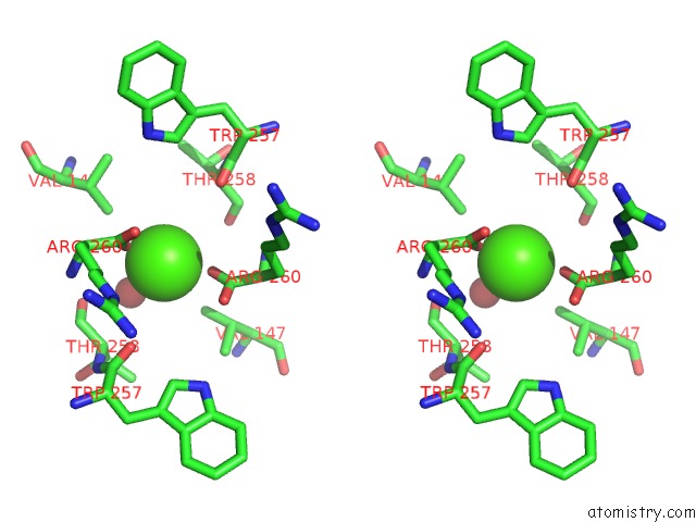
Calcium binding site 1 out of 2 in 3vdr
Go back to
Calcium binding site 1 out
of 2 in the Crystal Structure of D-3-Hydroxybutyrate Dehydrogenase, Prepared in the Presence of the Substrate D-3-Hydroxybutyrate and Nad(+)

Mono view

Stereo pair view
Mono view
Stereo pair view
A full contact list of Calcium with other atoms in the Ca binding
site number 1 of Crystal Structure of D-3-Hydroxybutyrate Dehydrogenase, Prepared in the Presence of the Substrate D-3-Hydroxybutyrate and Nad(+) within 5.0Å range:
|
Calcium binding site 2 out of 2 in 3vdr
Go back to
Calcium binding site 2 out
of 2 in the Crystal Structure of D-3-Hydroxybutyrate Dehydrogenase, Prepared in the Presence of the Substrate D-3-Hydroxybutyrate and Nad(+)

Mono view
Stereo pair view
Mono view
Stereo pair view
A full contact list of Calcium with other atoms in the Ca binding
site number 2 of Crystal Structure of D-3-Hydroxybutyrate Dehydrogenase, Prepared in the Presence of the Substrate D-3-Hydroxybutyrate and Nad(+) within 5.0Å range:
|
Reference:
M.M.Hoque,
S.Shimizu,
E.C.M.Juan,
Y.Sato,
M.T.Hossain,
T.Yamamoto,
S.Imamura,
K.Suzuki,
H.Amano,
T.Sekiguchi,
M.Tsunoda,
A.Takenaka.
Structure of D-3-Hydroxybutyrate Dehydrogenase Prepared in the Presence of the Substrate D-3-Hydroxybutyrate and Nad+. Acta Crystallogr.,Sect.F V. 65 331 2009.
ISSN: ESSN 1744-3091
PubMed: 19342772
DOI: 10.1107/S1744309109008537
Page generated: Tue Jul 8 17:34:05 2025
ISSN: ESSN 1744-3091
PubMed: 19342772
DOI: 10.1107/S1744309109008537
Last articles
Cl in 8BJKCl in 8BJE
Cl in 8BJD
Cl in 8BJG
Cl in 8BJ0
Cl in 8BIO
Cl in 8BIK
Cl in 8BIN
Cl in 8BIJ
Cl in 8BIH