Calcium »
PDB 3v0a-3vl2 »
3vj9 »
Calcium in PDB 3vj9: Crystal Structure of the Human Squalene Synthase
Enzymatic activity of Crystal Structure of the Human Squalene Synthase
All present enzymatic activity of Crystal Structure of the Human Squalene Synthase:
2.5.1.21;
2.5.1.21;
Protein crystallography data
The structure of Crystal Structure of the Human Squalene Synthase, PDB code: 3vj9
was solved by
C.I.Liu,
W.Y.Jeng,
W.J.Chang,
A.H.J.Wang,
with X-Ray Crystallography technique. A brief refinement statistics is given in the table below:
| Resolution Low / High (Å) | 24.70 / 1.52 |
| Space group | P 21 21 21 |
| Cell size a, b, c (Å), α, β, γ (°) | 51.776, 76.657, 82.501, 90.00, 90.00, 90.00 |
| R / Rfree (%) | 16 / 21.4 |
Other elements in 3vj9:
The structure of Crystal Structure of the Human Squalene Synthase also contains other interesting chemical elements:
| Nickel | (Ni) | 1 atom |
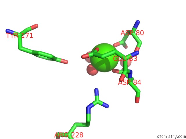
Calcium Binding Sites:
The binding sites of Calcium atom in the Crystal Structure of the Human Squalene Synthase
(pdb code 3vj9). This binding sites where shown within
5.0 Angstroms radius around Calcium atom.
In total only one binding site of Calcium was determined in the Crystal Structure of the Human Squalene Synthase, PDB code: 3vj9:
In total only one binding site of Calcium was determined in the Crystal Structure of the Human Squalene Synthase, PDB code: 3vj9:
Calcium binding site 1 out of 1 in 3vj9
Go back to
Calcium binding site 1 out
of 1 in the Crystal Structure of the Human Squalene Synthase
Mono view
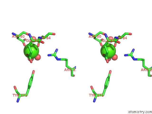
Stereo pair view
Mono view
Stereo pair view
A full contact list of Calcium with other atoms in the Ca binding
site number 1 of Crystal Structure of the Human Squalene Synthase within 5.0Å range:
|
Reference:
C.I.Liu,
W.Y.Jeng,
W.J.Chang,
T.P.Ko,
A.H.J.Wang.
Binding Modes of Zaragozic Acid A to Human Squalene Synthase and Staphylococcal Dehydrosqualene Synthase J.Biol.Chem. V. 287 18750 2012.
ISSN: ISSN 0021-9258
PubMed: 22474324
DOI: 10.1074/JBC.M112.351254
Page generated: Sat Jul 13 20:40:24 2024
ISSN: ISSN 0021-9258
PubMed: 22474324
DOI: 10.1074/JBC.M112.351254
Last articles
Zn in 9JYWZn in 9IR4
Zn in 9IR3
Zn in 9GMX
Zn in 9GMW
Zn in 9JEJ
Zn in 9ERF
Zn in 9ERE
Zn in 9EGV
Zn in 9EGW