Calcium »
PDB 3vl3-3vx1 »
3vlw »
Calcium in PDB 3vlw: Crystal Structure of Sphingomonas Sp. A1 Alginate-Binding Protein ALGQ1 in Complex with Mannuronate-Guluronate Disaccharide
Protein crystallography data
The structure of Crystal Structure of Sphingomonas Sp. A1 Alginate-Binding Protein ALGQ1 in Complex with Mannuronate-Guluronate Disaccharide, PDB code: 3vlw
was solved by
Y.Nishitani,
Y.Maruyama,
T.Itoh,
B.Mikami,
W.Hashimoto,
K.Murata,
with X-Ray Crystallography technique. A brief refinement statistics is given in the table below:
| Resolution Low / High (Å) | 50.00 / 2.00 |
| Space group | P 1 21 1 |
| Cell size a, b, c (Å), α, β, γ (°) | 79.759, 67.533, 90.693, 90.00, 93.33, 90.00 |
| R / Rfree (%) | 19 / 22.3 |
Calcium Binding Sites:
The binding sites of Calcium atom in the Crystal Structure of Sphingomonas Sp. A1 Alginate-Binding Protein ALGQ1 in Complex with Mannuronate-Guluronate Disaccharide
(pdb code 3vlw). This binding sites where shown within
5.0 Angstroms radius around Calcium atom.
In total 2 binding sites of Calcium where determined in the Crystal Structure of Sphingomonas Sp. A1 Alginate-Binding Protein ALGQ1 in Complex with Mannuronate-Guluronate Disaccharide, PDB code: 3vlw:
Jump to Calcium binding site number: 1; 2;
In total 2 binding sites of Calcium where determined in the Crystal Structure of Sphingomonas Sp. A1 Alginate-Binding Protein ALGQ1 in Complex with Mannuronate-Guluronate Disaccharide, PDB code: 3vlw:
Jump to Calcium binding site number: 1; 2;
Calcium binding site 1 out of 2 in 3vlw
Go back to
Calcium binding site 1 out
of 2 in the Crystal Structure of Sphingomonas Sp. A1 Alginate-Binding Protein ALGQ1 in Complex with Mannuronate-Guluronate Disaccharide
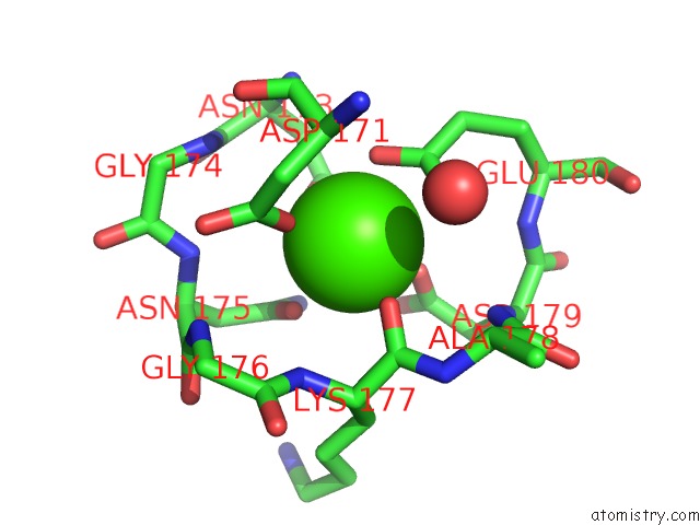
Mono view
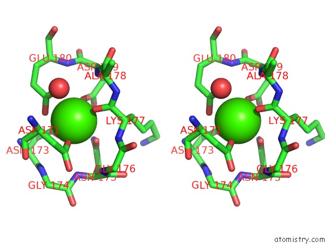
Stereo pair view
Mono view
Stereo pair view
A full contact list of Calcium with other atoms in the Ca binding
site number 1 of Crystal Structure of Sphingomonas Sp. A1 Alginate-Binding Protein ALGQ1 in Complex with Mannuronate-Guluronate Disaccharide within 5.0Å range:
|
Calcium binding site 2 out of 2 in 3vlw
Go back to
Calcium binding site 2 out
of 2 in the Crystal Structure of Sphingomonas Sp. A1 Alginate-Binding Protein ALGQ1 in Complex with Mannuronate-Guluronate Disaccharide
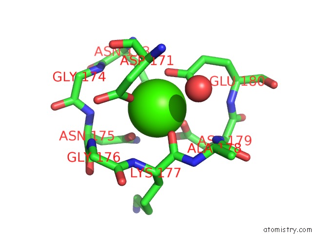
Mono view
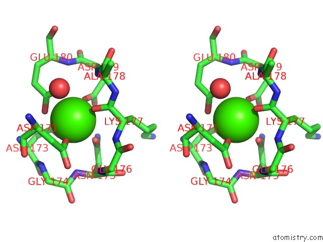
Stereo pair view
Mono view
Stereo pair view
A full contact list of Calcium with other atoms in the Ca binding
site number 2 of Crystal Structure of Sphingomonas Sp. A1 Alginate-Binding Protein ALGQ1 in Complex with Mannuronate-Guluronate Disaccharide within 5.0Å range:
|
Reference:
Y.Nishitani,
Y.Maruyama,
T.Itoh,
B.Mikami,
W.Hashimoto,
K.Murata.
Recognition of Heteropolysaccharide Alginate By Periplasmic Solute-Binding Proteins of A Bacterial Abc Transporter Biochemistry V. 51 3622 2012.
ISSN: ISSN 0006-2960
PubMed: 22486720
DOI: 10.1021/BI300194F
Page generated: Sat Jul 13 20:47:16 2024
ISSN: ISSN 0006-2960
PubMed: 22486720
DOI: 10.1021/BI300194F
Last articles
Zn in 9J0NZn in 9J0O
Zn in 9J0P
Zn in 9FJX
Zn in 9EKB
Zn in 9C0F
Zn in 9CAH
Zn in 9CH0
Zn in 9CH3
Zn in 9CH1