Calcium »
PDB 3vl3-3vx1 »
3vrq »
Calcium in PDB 3vrq: Crystal Structure of the Tyrosine Kinase Binding Domain of Cbl-C (Pl Mutant)
Protein crystallography data
The structure of Crystal Structure of the Tyrosine Kinase Binding Domain of Cbl-C (Pl Mutant), PDB code: 3vrq
was solved by
K.Takeshita,
T.Tezuka,
Y.Isozaki,
E.Yamashita,
M.Suzuki,
Y.Yamanashi,
T.Yamamoto,
A.Nakagawa,
with X-Ray Crystallography technique. A brief refinement statistics is given in the table below:
| Resolution Low / High (Å) | 37.98 / 2.39 |
| Space group | P 1 21 1 |
| Cell size a, b, c (Å), α, β, γ (°) | 62.600, 66.211, 81.558, 90.00, 106.02, 90.00 |
| R / Rfree (%) | 21.5 / 26.4 |
Calcium Binding Sites:
The binding sites of Calcium atom in the Crystal Structure of the Tyrosine Kinase Binding Domain of Cbl-C (Pl Mutant)
(pdb code 3vrq). This binding sites where shown within
5.0 Angstroms radius around Calcium atom.
In total 2 binding sites of Calcium where determined in the Crystal Structure of the Tyrosine Kinase Binding Domain of Cbl-C (Pl Mutant), PDB code: 3vrq:
Jump to Calcium binding site number: 1; 2;
In total 2 binding sites of Calcium where determined in the Crystal Structure of the Tyrosine Kinase Binding Domain of Cbl-C (Pl Mutant), PDB code: 3vrq:
Jump to Calcium binding site number: 1; 2;
Calcium binding site 1 out of 2 in 3vrq
Go back to
Calcium binding site 1 out
of 2 in the Crystal Structure of the Tyrosine Kinase Binding Domain of Cbl-C (Pl Mutant)
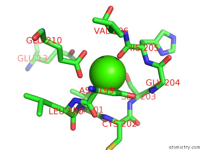
Mono view
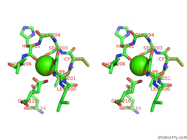
Stereo pair view
Mono view
Stereo pair view
A full contact list of Calcium with other atoms in the Ca binding
site number 1 of Crystal Structure of the Tyrosine Kinase Binding Domain of Cbl-C (Pl Mutant) within 5.0Å range:
|
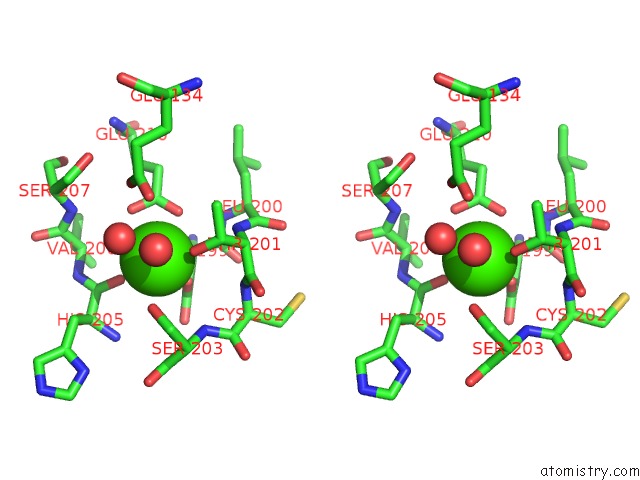
Calcium binding site 2 out of 2 in 3vrq
Go back to
Calcium binding site 2 out
of 2 in the Crystal Structure of the Tyrosine Kinase Binding Domain of Cbl-C (Pl Mutant)

Mono view
Stereo pair view
Mono view
Stereo pair view
A full contact list of Calcium with other atoms in the Ca binding
site number 2 of Crystal Structure of the Tyrosine Kinase Binding Domain of Cbl-C (Pl Mutant) within 5.0Å range:
|
Reference:
K.Takeshita,
T.Tezuka,
Y.Isozaki,
E.Yamashita,
M.Suzuki,
M.Kim,
Y.Yamanashi,
T.Yamamoto,
A.Nakagawa.
Structural Flexibility Regulates Phosphopeptide-Binding Activity of the Tyrosine Kinase Binding Domain of Cbl-C. J.Biochem. V. 152 487 2012.
ISSN: ISSN 0021-924X
PubMed: 22888118
DOI: 10.1093/JB/MVS085
Page generated: Sat Jul 13 20:49:43 2024
ISSN: ISSN 0021-924X
PubMed: 22888118
DOI: 10.1093/JB/MVS085
Last articles
Zn in 9J0NZn in 9J0O
Zn in 9J0P
Zn in 9FJX
Zn in 9EKB
Zn in 9C0F
Zn in 9CAH
Zn in 9CH0
Zn in 9CH3
Zn in 9CH1