Calcium »
PDB 3vyk-3whd »
3vyk »
Calcium in PDB 3vyk: Crystal Structure of C-Type Lectin Domain of Murine Dendritic Cell Inhibitory Receptor 2 in Complex with N-Glycan
Protein crystallography data
The structure of Crystal Structure of C-Type Lectin Domain of Murine Dendritic Cell Inhibitory Receptor 2 in Complex with N-Glycan, PDB code: 3vyk
was solved by
M.Nagae,
K.Yamanaka,
S.Hanashima,
A.Ikeda,
T.Satoh,
N.Matsumoto,
K.Yamamoto,
Y.Yamaguchi,
with X-Ray Crystallography technique. A brief refinement statistics is given in the table below:
| Resolution Low / High (Å) | 66.11 / 1.50 |
| Space group | P 32 2 1 |
| Cell size a, b, c (Å), α, β, γ (°) | 76.341, 76.341, 50.556, 90.00, 90.00, 120.00 |
| R / Rfree (%) | 21 / 22.4 |
Calcium Binding Sites:
The binding sites of Calcium atom in the Crystal Structure of C-Type Lectin Domain of Murine Dendritic Cell Inhibitory Receptor 2 in Complex with N-Glycan
(pdb code 3vyk). This binding sites where shown within
5.0 Angstroms radius around Calcium atom.
In total only one binding site of Calcium was determined in the Crystal Structure of C-Type Lectin Domain of Murine Dendritic Cell Inhibitory Receptor 2 in Complex with N-Glycan, PDB code: 3vyk:
In total only one binding site of Calcium was determined in the Crystal Structure of C-Type Lectin Domain of Murine Dendritic Cell Inhibitory Receptor 2 in Complex with N-Glycan, PDB code: 3vyk:
Calcium binding site 1 out of 1 in 3vyk
Go back to
Calcium binding site 1 out
of 1 in the Crystal Structure of C-Type Lectin Domain of Murine Dendritic Cell Inhibitory Receptor 2 in Complex with N-Glycan
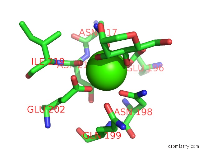
Mono view
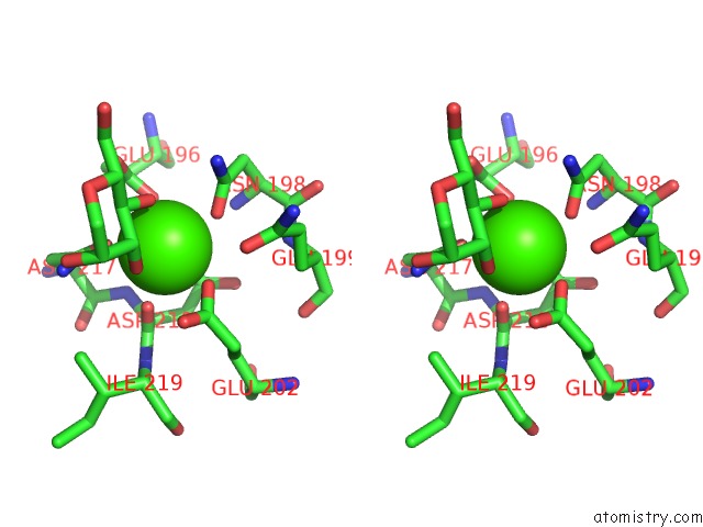
Stereo pair view
Mono view
Stereo pair view
A full contact list of Calcium with other atoms in the Ca binding
site number 1 of Crystal Structure of C-Type Lectin Domain of Murine Dendritic Cell Inhibitory Receptor 2 in Complex with N-Glycan within 5.0Å range:
|
Reference:
M.Nagae,
K.Yamanaka,
S.Hanashima,
A.Ikeda,
K.Morita-Matsumoto,
T.Satoh,
N.Matsumoto,
K.Yamamoto,
Y.Yamaguchi.
Recognition of Bisecting N-Acetylglucosamine: Structural Basis For Asymmetric Interaction with the Mouse Lectin Dendritic Cell Inhibitory Receptor 2 J.Biol.Chem. V. 288 33598 2013.
ISSN: ISSN 0021-9258
PubMed: 24108122
DOI: 10.1074/JBC.M113.513572
Page generated: Tue Jul 8 17:44:19 2025
ISSN: ISSN 0021-9258
PubMed: 24108122
DOI: 10.1074/JBC.M113.513572
Last articles
F in 7NEUF in 7NBW
F in 7NEC
F in 7NBL
F in 7N93
F in 7NBK
F in 7NAJ
F in 7N91
F in 7N8R
F in 7N77