Calcium »
PDB 3vyk-3whd »
3w5n »
Calcium in PDB 3w5n: Crystal Structure of Streptomyces Avermitilis Alpha-L-Rhamnosidase Complexed with L-Rhamnose
Enzymatic activity of Crystal Structure of Streptomyces Avermitilis Alpha-L-Rhamnosidase Complexed with L-Rhamnose
All present enzymatic activity of Crystal Structure of Streptomyces Avermitilis Alpha-L-Rhamnosidase Complexed with L-Rhamnose:
3.2.1.40;
3.2.1.40;
Protein crystallography data
The structure of Crystal Structure of Streptomyces Avermitilis Alpha-L-Rhamnosidase Complexed with L-Rhamnose, PDB code: 3w5n
was solved by
Z.Fujimoto,
A.Jackson,
M.Michikawa,
T.Maehara,
M.Momma,
B.F.Henrissat,
H.J.Gilbert,
S.Kaneko,
with X-Ray Crystallography technique. A brief refinement statistics is given in the table below:
| Resolution Low / High (Å) | 40.56 / 1.80 |
| Space group | P 1 21 1 |
| Cell size a, b, c (Å), α, β, γ (°) | 53.063, 128.559, 75.260, 90.00, 99.85, 90.00 |
| R / Rfree (%) | 16.3 / 19.4 |
Other elements in 3w5n:
The structure of Crystal Structure of Streptomyces Avermitilis Alpha-L-Rhamnosidase Complexed with L-Rhamnose also contains other interesting chemical elements:
| Sodium | (Na) | 3 atoms |
Calcium Binding Sites:
The binding sites of Calcium atom in the Crystal Structure of Streptomyces Avermitilis Alpha-L-Rhamnosidase Complexed with L-Rhamnose
(pdb code 3w5n). This binding sites where shown within
5.0 Angstroms radius around Calcium atom.
In total only one binding site of Calcium was determined in the Crystal Structure of Streptomyces Avermitilis Alpha-L-Rhamnosidase Complexed with L-Rhamnose, PDB code: 3w5n:
In total only one binding site of Calcium was determined in the Crystal Structure of Streptomyces Avermitilis Alpha-L-Rhamnosidase Complexed with L-Rhamnose, PDB code: 3w5n:
Calcium binding site 1 out of 1 in 3w5n
Go back to
Calcium binding site 1 out
of 1 in the Crystal Structure of Streptomyces Avermitilis Alpha-L-Rhamnosidase Complexed with L-Rhamnose
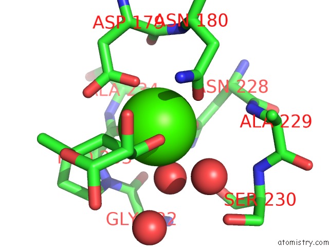
Mono view
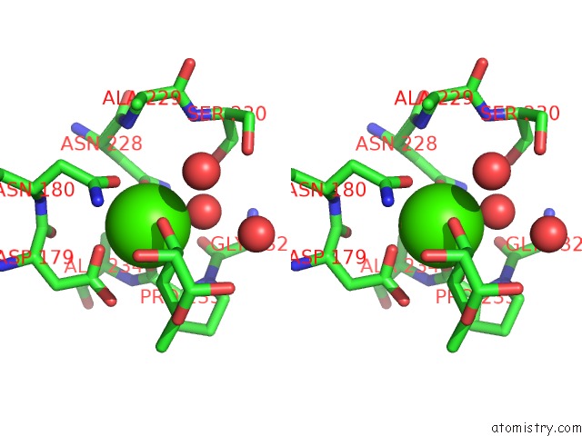
Stereo pair view
Mono view
Stereo pair view
A full contact list of Calcium with other atoms in the Ca binding
site number 1 of Crystal Structure of Streptomyces Avermitilis Alpha-L-Rhamnosidase Complexed with L-Rhamnose within 5.0Å range:
|
Reference:
Z.Fujimoto,
A.Jackson,
M.Michikawa,
T.Maehara,
M.Momma,
B.F.Henrissat,
H.J.Gilbert,
S.Kaneko.
The Structure of A Streptomyces Avermitilis Alpha-L-Rhamnosidase Reveals A Novel Carbohydrate-Binding Module CBM67 Within the Six-Domain Arrangement. J.Biol.Chem. V. 288 12376 2013.
ISSN: ISSN 0021-9258
PubMed: 23486481
DOI: 10.1074/JBC.M113.460097
Page generated: Sat Jul 13 20:55:28 2024
ISSN: ISSN 0021-9258
PubMed: 23486481
DOI: 10.1074/JBC.M113.460097
Last articles
Zn in 9J0NZn in 9J0O
Zn in 9J0P
Zn in 9FJX
Zn in 9EKB
Zn in 9C0F
Zn in 9CAH
Zn in 9CH0
Zn in 9CH3
Zn in 9CH1