Calcium »
PDB 3vyk-3whd »
3w7u »
Calcium in PDB 3w7u: Escherichia Coli K12 Ygjk Complexed with Galactose
Protein crystallography data
The structure of Escherichia Coli K12 Ygjk Complexed with Galactose, PDB code: 3w7u
was solved by
T.Miyazaki,
Y.Kurakata,
A.Uechi,
H.Yoshida,
S.Kamitori,
Y.Sakano,
A.Nishikawa,
T.Tonozuka,
with X-Ray Crystallography technique. A brief refinement statistics is given in the table below:
| Resolution Low / High (Å) | 36.25 / 1.99 |
| Space group | P 1 21 1 |
| Cell size a, b, c (Å), α, β, γ (°) | 62.070, 140.020, 86.260, 90.00, 97.66, 90.00 |
| R / Rfree (%) | 19.1 / 23.6 |
Calcium Binding Sites:
The binding sites of Calcium atom in the Escherichia Coli K12 Ygjk Complexed with Galactose
(pdb code 3w7u). This binding sites where shown within
5.0 Angstroms radius around Calcium atom.
In total 2 binding sites of Calcium where determined in the Escherichia Coli K12 Ygjk Complexed with Galactose, PDB code: 3w7u:
Jump to Calcium binding site number: 1; 2;
In total 2 binding sites of Calcium where determined in the Escherichia Coli K12 Ygjk Complexed with Galactose, PDB code: 3w7u:
Jump to Calcium binding site number: 1; 2;
Calcium binding site 1 out of 2 in 3w7u
Go back to
Calcium binding site 1 out
of 2 in the Escherichia Coli K12 Ygjk Complexed with Galactose
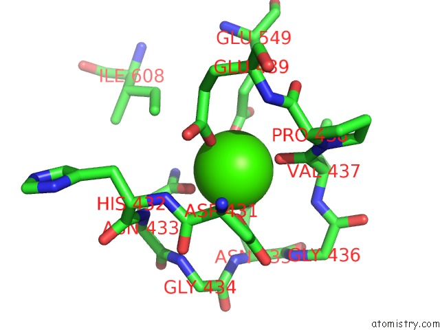
Mono view
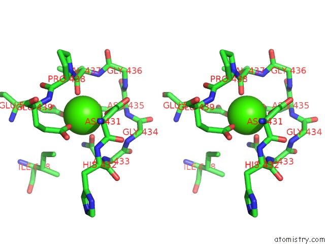
Stereo pair view
Mono view
Stereo pair view
A full contact list of Calcium with other atoms in the Ca binding
site number 1 of Escherichia Coli K12 Ygjk Complexed with Galactose within 5.0Å range:
|
Calcium binding site 2 out of 2 in 3w7u
Go back to
Calcium binding site 2 out
of 2 in the Escherichia Coli K12 Ygjk Complexed with Galactose
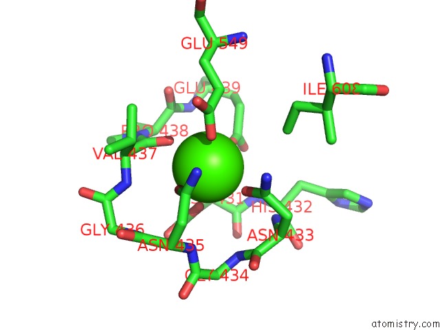
Mono view
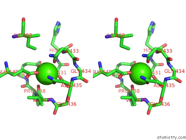
Stereo pair view
Mono view
Stereo pair view
A full contact list of Calcium with other atoms in the Ca binding
site number 2 of Escherichia Coli K12 Ygjk Complexed with Galactose within 5.0Å range:
|
Reference:
Y.Kurakata,
A.Uechi,
H.Yoshida,
S.Kamitori,
Y.Sakano,
A.Nishikawa,
T.Tonozuka.
Structural Insights Into the Substrate Specificity and Function of Escherichia Coli K12 Ygjk, A Glucosidase Belonging to the Glycoside Hydrolase Family 63. J.Mol.Biol. V. 381 116 2008.
ISSN: ISSN 0022-2836
PubMed: 18586271
DOI: 10.1016/J.JMB.2008.05.061
Page generated: Sat Jul 13 20:56:33 2024
ISSN: ISSN 0022-2836
PubMed: 18586271
DOI: 10.1016/J.JMB.2008.05.061
Last articles
Zn in 9J0NZn in 9J0O
Zn in 9J0P
Zn in 9FJX
Zn in 9EKB
Zn in 9C0F
Zn in 9CAH
Zn in 9CH0
Zn in 9CH3
Zn in 9CH1