Calcium »
PDB 3vyk-3whd »
3w7x »
Calcium in PDB 3w7x: Crystal Structure of E. Coli Ygjk D324N Complexed with Melibiose
Protein crystallography data
The structure of Crystal Structure of E. Coli Ygjk D324N Complexed with Melibiose, PDB code: 3w7x
was solved by
T.Miyazaki,
M.Ichikawa,
G.Yokoi,
M.Kitaoka,
H.Mori,
Y.Kitano,
A.Nishikawa,
T.Tonozuka,
with X-Ray Crystallography technique. A brief refinement statistics is given in the table below:
| Resolution Low / High (Å) | 40.52 / 2.70 |
| Space group | P 1 21 1 |
| Cell size a, b, c (Å), α, β, γ (°) | 62.140, 138.160, 86.200, 90.00, 98.37, 90.00 |
| R / Rfree (%) | 18.9 / 24.9 |
Calcium Binding Sites:
The binding sites of Calcium atom in the Crystal Structure of E. Coli Ygjk D324N Complexed with Melibiose
(pdb code 3w7x). This binding sites where shown within
5.0 Angstroms radius around Calcium atom.
In total 2 binding sites of Calcium where determined in the Crystal Structure of E. Coli Ygjk D324N Complexed with Melibiose, PDB code: 3w7x:
Jump to Calcium binding site number: 1; 2;
In total 2 binding sites of Calcium where determined in the Crystal Structure of E. Coli Ygjk D324N Complexed with Melibiose, PDB code: 3w7x:
Jump to Calcium binding site number: 1; 2;
Calcium binding site 1 out of 2 in 3w7x
Go back to
Calcium binding site 1 out
of 2 in the Crystal Structure of E. Coli Ygjk D324N Complexed with Melibiose
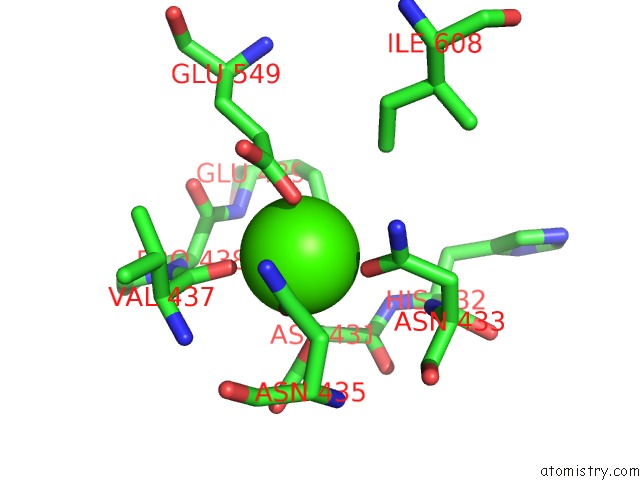
Mono view
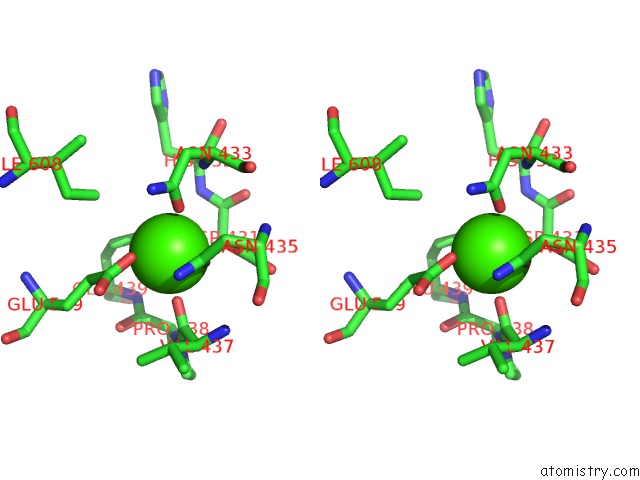
Stereo pair view
Mono view
Stereo pair view
A full contact list of Calcium with other atoms in the Ca binding
site number 1 of Crystal Structure of E. Coli Ygjk D324N Complexed with Melibiose within 5.0Å range:
|
Calcium binding site 2 out of 2 in 3w7x
Go back to
Calcium binding site 2 out
of 2 in the Crystal Structure of E. Coli Ygjk D324N Complexed with Melibiose
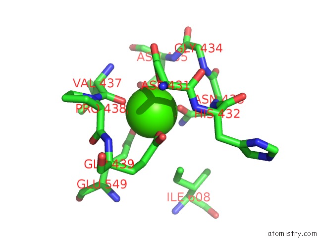
Mono view
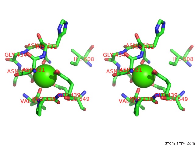
Stereo pair view
Mono view
Stereo pair view
A full contact list of Calcium with other atoms in the Ca binding
site number 2 of Crystal Structure of E. Coli Ygjk D324N Complexed with Melibiose within 5.0Å range:
|
Reference:
T.Miyazaki,
M.Ichikawa,
G.Yokoi,
M.Kitaoka,
H.Mori,
Y.Kitano,
A.Nishikawa,
T.Tonozuka.
Structure of A Bacterial Glycoside Hydrolase Family 63 Enzyme in Complex with Its Glycosynthase Product, and Insights Into the Substrate Specificity. Febs J. V. 280 4560 2013.
ISSN: ISSN 1742-464X
PubMed: 23826932
DOI: 10.1111/FEBS.12424
Page generated: Sat Jul 13 20:57:53 2024
ISSN: ISSN 1742-464X
PubMed: 23826932
DOI: 10.1111/FEBS.12424
Last articles
Zn in 9JYWZn in 9IR4
Zn in 9IR3
Zn in 9GMX
Zn in 9GMW
Zn in 9JEJ
Zn in 9ERF
Zn in 9ERE
Zn in 9EGV
Zn in 9EGW