Calcium »
PDB 3whi-3ws5 »
3ws5 »
Calcium in PDB 3ws5: N288Q-N321Q Mutant Beta-Lactamase Derived From Chromohalobacter Sp.560 (Condition-2B)
Enzymatic activity of N288Q-N321Q Mutant Beta-Lactamase Derived From Chromohalobacter Sp.560 (Condition-2B)
All present enzymatic activity of N288Q-N321Q Mutant Beta-Lactamase Derived From Chromohalobacter Sp.560 (Condition-2B):
3.5.2.6;
3.5.2.6;
Protein crystallography data
The structure of N288Q-N321Q Mutant Beta-Lactamase Derived From Chromohalobacter Sp.560 (Condition-2B), PDB code: 3ws5
was solved by
S.Arai,
Y.Yonezawa,
N.Okazaki,
F.Matsumoto,
R.Shimizu,
M.Yamada,
M.Adachi,
T.Tamada,
H.Tokunaga,
M.Ishibashi,
M.Tokunaga,
R.Kuroki,
with X-Ray Crystallography technique. A brief refinement statistics is given in the table below:
| Resolution Low / High (Å) | 29.59 / 2.80 |
| Space group | P 31 |
| Cell size a, b, c (Å), α, β, γ (°) | 114.262, 114.262, 66.998, 90.00, 90.00, 120.00 |
| R / Rfree (%) | 17.6 / 20.7 |
Other elements in 3ws5:
The structure of N288Q-N321Q Mutant Beta-Lactamase Derived From Chromohalobacter Sp.560 (Condition-2B) also contains other interesting chemical elements:
| Strontium | (Sr) | 1 atom |
| Chlorine | (Cl) | 3 atoms |
Calcium Binding Sites:
The binding sites of Calcium atom in the N288Q-N321Q Mutant Beta-Lactamase Derived From Chromohalobacter Sp.560 (Condition-2B)
(pdb code 3ws5). This binding sites where shown within
5.0 Angstroms radius around Calcium atom.
In total only one binding site of Calcium was determined in the N288Q-N321Q Mutant Beta-Lactamase Derived From Chromohalobacter Sp.560 (Condition-2B), PDB code: 3ws5:
In total only one binding site of Calcium was determined in the N288Q-N321Q Mutant Beta-Lactamase Derived From Chromohalobacter Sp.560 (Condition-2B), PDB code: 3ws5:
Calcium binding site 1 out of 1 in 3ws5
Go back to
Calcium binding site 1 out
of 1 in the N288Q-N321Q Mutant Beta-Lactamase Derived From Chromohalobacter Sp.560 (Condition-2B)
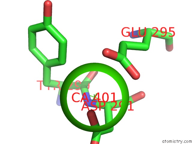
Mono view
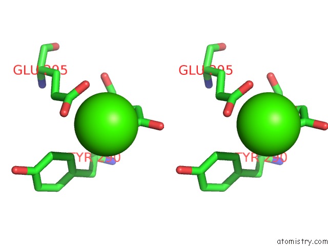
Stereo pair view
Mono view
Stereo pair view
A full contact list of Calcium with other atoms in the Ca binding
site number 1 of N288Q-N321Q Mutant Beta-Lactamase Derived From Chromohalobacter Sp.560 (Condition-2B) within 5.0Å range:
|
Reference:
S.Arai,
Y.Yonezawa,
N.Okazaki,
F.Matsumoto,
C.Shibazaki,
R.Shimizu,
M.Yamada,
M.Adachi,
T.Tamada,
T.Kawamoto,
H.Tokunaga,
M.Ishibashi,
M.Blaber,
M.Tokunaga,
R.Kuroki.
Crystal Structure of Highly Acidic Beta-Lactamase From Moderate Halophile Chromohalobacter Sp. 560 and the Discovery of A Cs+ Selective Binding Site To Be Published.
Page generated: Sat Jul 13 21:21:58 2024
Last articles
Zn in 9J0NZn in 9J0O
Zn in 9J0P
Zn in 9FJX
Zn in 9EKB
Zn in 9C0F
Zn in 9CAH
Zn in 9CH0
Zn in 9CH3
Zn in 9CH1