Calcium »
PDB 3wt3-3zq4 »
3zoa »
Calcium in PDB 3zoa: The Structure of Trehalose Synthase (Tres) of Mycobacterium Smegmatis in Complex with Acarbose
Enzymatic activity of The Structure of Trehalose Synthase (Tres) of Mycobacterium Smegmatis in Complex with Acarbose
All present enzymatic activity of The Structure of Trehalose Synthase (Tres) of Mycobacterium Smegmatis in Complex with Acarbose:
3.2.1.1; 5.4.99.16;
3.2.1.1; 5.4.99.16;
Protein crystallography data
The structure of The Structure of Trehalose Synthase (Tres) of Mycobacterium Smegmatis in Complex with Acarbose, PDB code: 3zoa
was solved by
S.Caner,
N.Nguyen,
A.Aguda,
R.Zhang,
Y.T.Pan,
S.G.Withers,
G.D.Brayer,
with X-Ray Crystallography technique. A brief refinement statistics is given in the table below:
| Resolution Low / High (Å) | 34.816 / 1.85 |
| Space group | I 41 |
| Cell size a, b, c (Å), α, β, γ (°) | 127.180, 127.180, 216.950, 90.00, 90.00, 90.00 |
| R / Rfree (%) | 16.5 / 19.81 |
Other elements in 3zoa:
The structure of The Structure of Trehalose Synthase (Tres) of Mycobacterium Smegmatis in Complex with Acarbose also contains other interesting chemical elements:
| Magnesium | (Mg) | 2 atoms |
| Chlorine | (Cl) | 7 atoms |
Calcium Binding Sites:
The binding sites of Calcium atom in the The Structure of Trehalose Synthase (Tres) of Mycobacterium Smegmatis in Complex with Acarbose
(pdb code 3zoa). This binding sites where shown within
5.0 Angstroms radius around Calcium atom.
In total 2 binding sites of Calcium where determined in the The Structure of Trehalose Synthase (Tres) of Mycobacterium Smegmatis in Complex with Acarbose, PDB code: 3zoa:
Jump to Calcium binding site number: 1; 2;
In total 2 binding sites of Calcium where determined in the The Structure of Trehalose Synthase (Tres) of Mycobacterium Smegmatis in Complex with Acarbose, PDB code: 3zoa:
Jump to Calcium binding site number: 1; 2;
Calcium binding site 1 out of 2 in 3zoa
Go back to
Calcium binding site 1 out
of 2 in the The Structure of Trehalose Synthase (Tres) of Mycobacterium Smegmatis in Complex with Acarbose
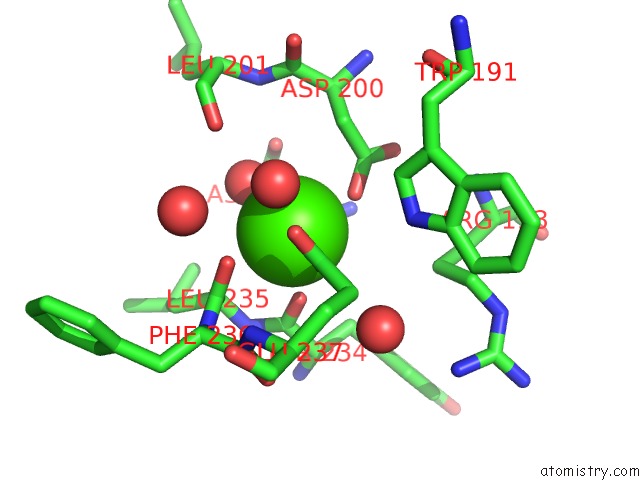
Mono view
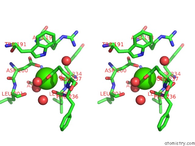
Stereo pair view
Mono view
Stereo pair view
A full contact list of Calcium with other atoms in the Ca binding
site number 1 of The Structure of Trehalose Synthase (Tres) of Mycobacterium Smegmatis in Complex with Acarbose within 5.0Å range:
|
Calcium binding site 2 out of 2 in 3zoa
Go back to
Calcium binding site 2 out
of 2 in the The Structure of Trehalose Synthase (Tres) of Mycobacterium Smegmatis in Complex with Acarbose
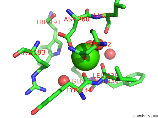
Mono view
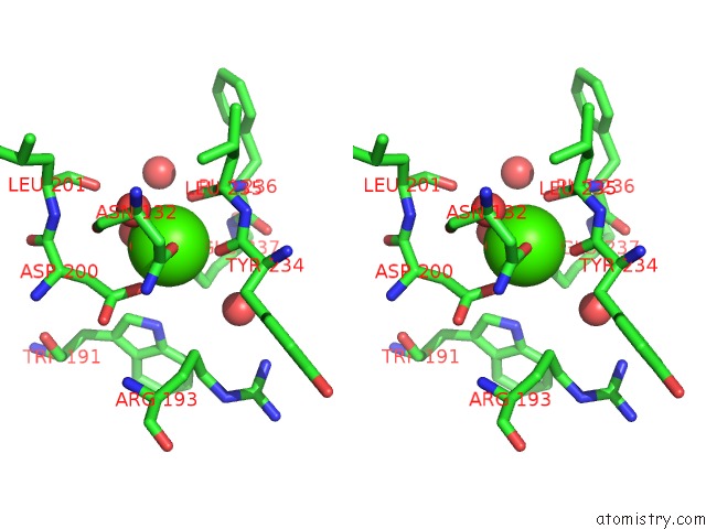
Stereo pair view
Mono view
Stereo pair view
A full contact list of Calcium with other atoms in the Ca binding
site number 2 of The Structure of Trehalose Synthase (Tres) of Mycobacterium Smegmatis in Complex with Acarbose within 5.0Å range:
|
Reference:
S.Caner,
N.Nguyen,
A.Aguda,
R.Zhang,
Y.T.Pan,
S.G.Withers,
G.D.Brayer.
The Structure of the Mycobacterium Smegmatis Trehalose Synthase Reveals An Unusual Active Site Configuration and Acarbose-Binding Mode. Glycobiology V. 23 1075 2013.
ISSN: ISSN 0959-6658
PubMed: 23735230
DOI: 10.1093/GLYCOB/CWT044
Page generated: Sat Jul 13 21:42:11 2024
ISSN: ISSN 0959-6658
PubMed: 23735230
DOI: 10.1093/GLYCOB/CWT044
Last articles
Zn in 9J0NZn in 9J0O
Zn in 9J0P
Zn in 9FJX
Zn in 9EKB
Zn in 9C0F
Zn in 9CAH
Zn in 9CH0
Zn in 9CH3
Zn in 9CH1