Calcium »
PDB 3zq9-4a41 »
3zyp »
Calcium in PDB 3zyp: Cellulose Induced Protein, CIP1
Protein crystallography data
The structure of Cellulose Induced Protein, CIP1, PDB code: 3zyp
was solved by
F.Jacobson,
S.Karkehabadi,
H.Hansson,
F.Goedegebuur,
L.Wallace,
C.Mitchinson,
K.Piens,
I.Stals,
M.Sandgren,
with X-Ray Crystallography technique. A brief refinement statistics is given in the table below:
| Resolution Low / High (Å) | 45.64 / 1.50 |
| Space group | P 21 21 21 |
| Cell size a, b, c (Å), α, β, γ (°) | 55.413, 57.515, 74.579, 90.00, 90.00, 90.00 |
| R / Rfree (%) | 19.115 / 21.668 |
Calcium Binding Sites:
The binding sites of Calcium atom in the Cellulose Induced Protein, CIP1
(pdb code 3zyp). This binding sites where shown within
5.0 Angstroms radius around Calcium atom.
In total only one binding site of Calcium was determined in the Cellulose Induced Protein, CIP1, PDB code: 3zyp:
In total only one binding site of Calcium was determined in the Cellulose Induced Protein, CIP1, PDB code: 3zyp:
Calcium binding site 1 out of 1 in 3zyp
Go back to
Calcium binding site 1 out
of 1 in the Cellulose Induced Protein, CIP1
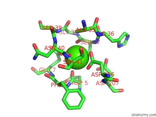
Mono view
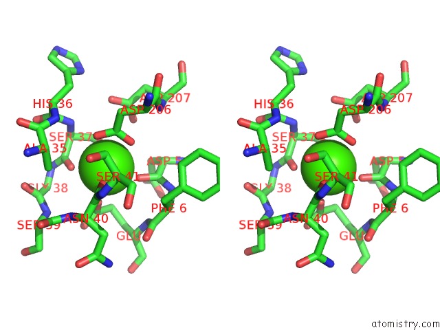
Stereo pair view
Mono view
Stereo pair view
A full contact list of Calcium with other atoms in the Ca binding
site number 1 of Cellulose Induced Protein, CIP1 within 5.0Å range:
|
Reference:
F.Jacobson,
S.Karkehabadi,
H.Hansson,
F.Goedegebuur,
L.Wallace,
C.Mitchinson,
K.Piens,
I.Stals,
M.Sandgren.
The Crystal Structure of the Core Domain of A Cellulose Induced Protein (CIP1) From Hypocrea Jecorina, at 1.5 A Resolution. Plos One V. 8 70562 2013.
ISSN: ISSN 1932-6203
PubMed: 24039705
DOI: 10.1371/JOURNAL.PONE.0070562
Page generated: Tue Jul 8 18:21:37 2025
ISSN: ISSN 1932-6203
PubMed: 24039705
DOI: 10.1371/JOURNAL.PONE.0070562
Last articles
Ca in 4L9WCa in 4L9O
Ca in 4L9S
Ca in 4L75
Ca in 4L9D
Ca in 4L73
Ca in 4L8Q
Ca in 4L7V
Ca in 4L74
Ca in 4L3Z