Calcium »
PDB 3zq9-4a41 »
4a0a »
Calcium in PDB 4a0a: Structure of HSDDB1-DRDDB2 Bound to A 16 Bp Cpd-Duplex (Pyrimidine at D-1 Position) at 3.6 A Resolution (Cpd 3)
Protein crystallography data
The structure of Structure of HSDDB1-DRDDB2 Bound to A 16 Bp Cpd-Duplex (Pyrimidine at D-1 Position) at 3.6 A Resolution (Cpd 3), PDB code: 4a0a
was solved by
A.Scrima,
E.S.Fischer,
S.Iwai,
H.Gut,
N.H.Thoma,
with X-Ray Crystallography technique. A brief refinement statistics is given in the table below:
| Resolution Low / High (Å) | 46.25 / 3.60 |
| Space group | C 2 2 21 |
| Cell size a, b, c (Å), α, β, γ (°) | 155.640, 227.140, 114.320, 90.00, 90.00, 90.00 |
| R / Rfree (%) | 26.3 / 34.7 |
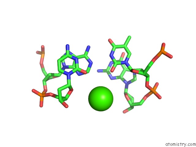
Calcium Binding Sites:
The binding sites of Calcium atom in the Structure of HSDDB1-DRDDB2 Bound to A 16 Bp Cpd-Duplex (Pyrimidine at D-1 Position) at 3.6 A Resolution (Cpd 3)
(pdb code 4a0a). This binding sites where shown within
5.0 Angstroms radius around Calcium atom.
In total only one binding site of Calcium was determined in the Structure of HSDDB1-DRDDB2 Bound to A 16 Bp Cpd-Duplex (Pyrimidine at D-1 Position) at 3.6 A Resolution (Cpd 3), PDB code: 4a0a:
In total only one binding site of Calcium was determined in the Structure of HSDDB1-DRDDB2 Bound to A 16 Bp Cpd-Duplex (Pyrimidine at D-1 Position) at 3.6 A Resolution (Cpd 3), PDB code: 4a0a:
Calcium binding site 1 out of 1 in 4a0a
Go back to
Calcium binding site 1 out
of 1 in the Structure of HSDDB1-DRDDB2 Bound to A 16 Bp Cpd-Duplex (Pyrimidine at D-1 Position) at 3.6 A Resolution (Cpd 3)
Mono view
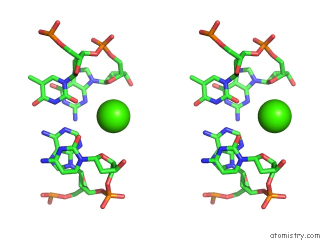
Stereo pair view
Mono view
Stereo pair view
A full contact list of Calcium with other atoms in the Ca binding
site number 1 of Structure of HSDDB1-DRDDB2 Bound to A 16 Bp Cpd-Duplex (Pyrimidine at D-1 Position) at 3.6 A Resolution (Cpd 3) within 5.0Å range:
|
Reference:
A.Scrima,
E.S.Fischer,
S.Iwai,
H.Gut,
N.H.Thoma.
The Molecular Basis of CRL4(DDB2/Csa) Ubiquitin Ligase Architecture, Targeting, and Activation Cell(Cambridge,Mass.) V. 147 1024 2011.
ISSN: ISSN 0092-8674
PubMed: 22118460
DOI: 10.1016/J.CELL.2011.10.035
Page generated: Sat Jul 13 21:53:11 2024
ISSN: ISSN 0092-8674
PubMed: 22118460
DOI: 10.1016/J.CELL.2011.10.035
Last articles
Zn in 9J0NZn in 9J0O
Zn in 9J0P
Zn in 9FJX
Zn in 9EKB
Zn in 9C0F
Zn in 9CAH
Zn in 9CH0
Zn in 9CH3
Zn in 9CH1