Calcium »
PDB 4a42-4adj »
4a5w »
Calcium in PDB 4a5w: Crystal Structure of C5B6
Protein crystallography data
The structure of Crystal Structure of C5B6, PDB code: 4a5w
was solved by
M.A.Hadders,
D.Bubeck,
F.Forneris,
M.Pangburn,
O.Llorca,
S.M.Lea,
P.Gros,
with X-Ray Crystallography technique. A brief refinement statistics is given in the table below:
| Resolution Low / High (Å) | 46.19 / 3.50 |
| Space group | I 21 21 21 |
| Cell size a, b, c (Å), α, β, γ (°) | 154.217, 230.747, 269.983, 90.00, 90.00, 90.00 |
| R / Rfree (%) | 25.6 / 27 |
Calcium Binding Sites:
The binding sites of Calcium atom in the Crystal Structure of C5B6
(pdb code 4a5w). This binding sites where shown within
5.0 Angstroms radius around Calcium atom.
In total only one binding site of Calcium was determined in the Crystal Structure of C5B6, PDB code: 4a5w:
In total only one binding site of Calcium was determined in the Crystal Structure of C5B6, PDB code: 4a5w:
Calcium binding site 1 out of 1 in 4a5w
Go back to
Calcium binding site 1 out
of 1 in the Crystal Structure of C5B6
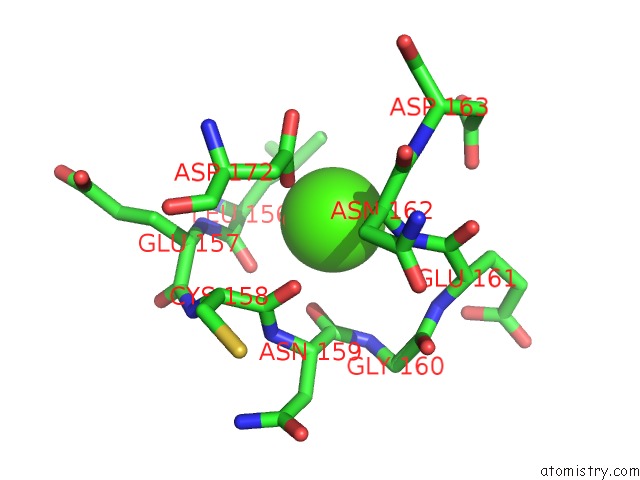
Mono view
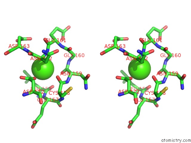
Stereo pair view
Mono view
Stereo pair view
A full contact list of Calcium with other atoms in the Ca binding
site number 1 of Crystal Structure of C5B6 within 5.0Å range:
|
Reference:
M.A.Hadders,
D.Bubeck,
P.Roversi,
S.Hakobyan,
F.Forneris,
B.P.Morgan,
M.K.Pangburn,
O.Llorca,
S.M.Lea,
P.Gros.
Assembly and Regulation of the Membrane Attack Complex Based on Structures of C5B6 and SC5B9. Cell Rep. V. 1 200 2012.
ISSN: ESSN 2211-1247
PubMed: 22832194
DOI: 10.1016/J.CELREP.2012.02.003
Page generated: Tue Jul 8 18:24:51 2025
ISSN: ESSN 2211-1247
PubMed: 22832194
DOI: 10.1016/J.CELREP.2012.02.003
Last articles
Fe in 2YXOFe in 2YRS
Fe in 2YXC
Fe in 2YNM
Fe in 2YVJ
Fe in 2YP1
Fe in 2YU2
Fe in 2YU1
Fe in 2YQB
Fe in 2YOO