Calcium in PDB 4a7h: Structure of the Actin-Tropomyosin-Myosin Complex (Rigor Atm 2)
Enzymatic activity of Structure of the Actin-Tropomyosin-Myosin Complex (Rigor Atm 2)
All present enzymatic activity of Structure of the Actin-Tropomyosin-Myosin Complex (Rigor Atm 2):
3.6.4.1;
Calcium Binding Sites:
The binding sites of Calcium atom in the Structure of the Actin-Tropomyosin-Myosin Complex (Rigor Atm 2)
(pdb code 4a7h). This binding sites where shown within
5.0 Angstroms radius around Calcium atom.
In total 5 binding sites of Calcium where determined in the
Structure of the Actin-Tropomyosin-Myosin Complex (Rigor Atm 2), PDB code: 4a7h:
Jump to Calcium binding site number:
1;
2;
3;
4;
5;
Calcium binding site 1 out
of 5 in 4a7h
Go back to
Calcium Binding Sites List in 4a7h
Calcium binding site 1 out
of 5 in the Structure of the Actin-Tropomyosin-Myosin Complex (Rigor Atm 2)
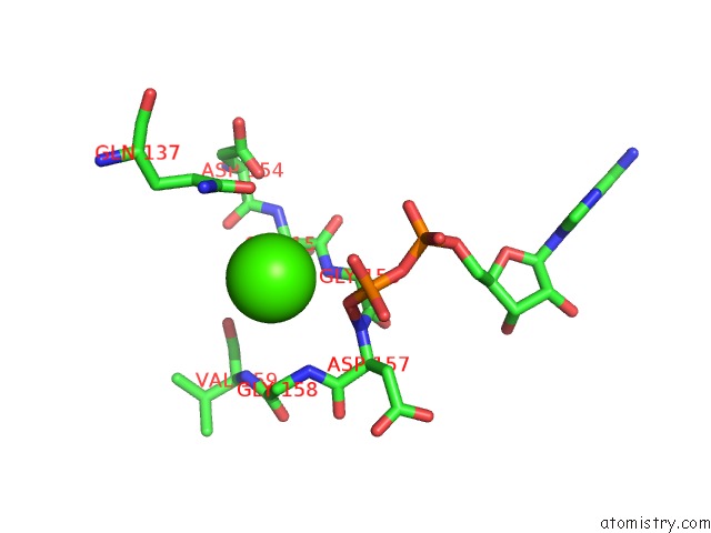
Mono view
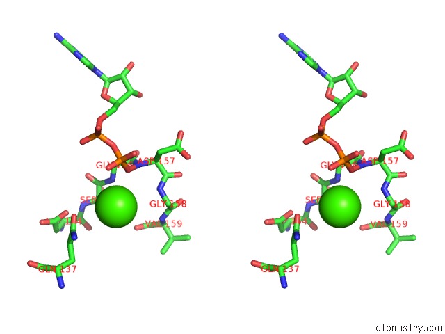
Stereo pair view
|
A full contact list of Calcium with other atoms in the Ca binding
site number 1 of Structure of the Actin-Tropomyosin-Myosin Complex (Rigor Atm 2) within 5.0Å range:
probe
|
atom
|
residue
|
distance (Å)
|
B
|
Occ
|
A:Ca377
b:12.4
occ:1.00
|
O3B
|
A:ADP376
|
3.3
|
10.0
|
1.0
|
N
|
A:GLY156
|
3.5
|
0.0
|
1.0
|
OE1
|
A:GLN137
|
3.8
|
0.0
|
1.0
|
C
|
A:SER155
|
4.0
|
0.0
|
1.0
|
N
|
A:GLY158
|
4.0
|
0.0
|
1.0
|
CA
|
A:GLY156
|
4.1
|
0.0
|
1.0
|
O1B
|
A:ADP376
|
4.1
|
11.2
|
1.0
|
CA
|
A:SER155
|
4.1
|
0.0
|
1.0
|
N
|
A:ASP157
|
4.2
|
0.0
|
1.0
|
PB
|
A:ADP376
|
4.3
|
10.8
|
1.0
|
CD
|
A:GLN137
|
4.4
|
0.0
|
1.0
|
CA
|
A:GLY158
|
4.5
|
0.0
|
1.0
|
C
|
A:GLY156
|
4.6
|
0.0
|
1.0
|
OD1
|
A:ASP154
|
4.7
|
0.0
|
1.0
|
O
|
A:VAL159
|
4.8
|
0.0
|
1.0
|
O
|
A:SER155
|
4.8
|
0.0
|
1.0
|
N
|
A:VAL159
|
4.9
|
0.0
|
1.0
|
CG
|
A:GLN137
|
5.0
|
0.0
|
1.0
|
N
|
A:SER155
|
5.0
|
0.0
|
1.0
|
NE2
|
A:GLN137
|
5.0
|
0.0
|
1.0
|
|
Calcium binding site 2 out
of 5 in 4a7h
Go back to
Calcium Binding Sites List in 4a7h
Calcium binding site 2 out
of 5 in the Structure of the Actin-Tropomyosin-Myosin Complex (Rigor Atm 2)
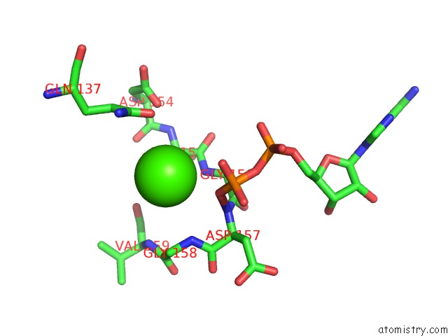
Mono view
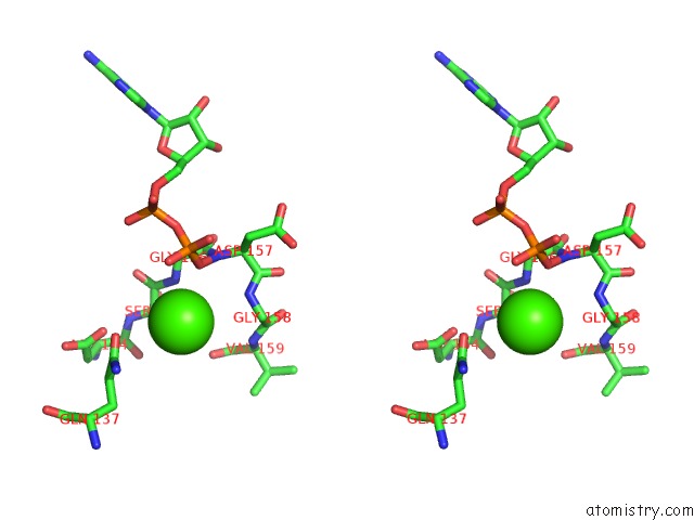
Stereo pair view
|
A full contact list of Calcium with other atoms in the Ca binding
site number 2 of Structure of the Actin-Tropomyosin-Myosin Complex (Rigor Atm 2) within 5.0Å range:
probe
|
atom
|
residue
|
distance (Å)
|
B
|
Occ
|
D:Ca377
b:12.4
occ:1.00
|
O3B
|
D:ADP376
|
3.3
|
10.0
|
1.0
|
N
|
D:GLY156
|
3.5
|
0.0
|
1.0
|
OE1
|
D:GLN137
|
3.8
|
0.0
|
1.0
|
C
|
D:SER155
|
4.0
|
0.0
|
1.0
|
N
|
D:GLY158
|
4.0
|
0.0
|
1.0
|
CA
|
D:GLY156
|
4.1
|
0.0
|
1.0
|
O1B
|
D:ADP376
|
4.1
|
11.2
|
1.0
|
CA
|
D:SER155
|
4.1
|
0.0
|
1.0
|
N
|
D:ASP157
|
4.2
|
0.0
|
1.0
|
PB
|
D:ADP376
|
4.3
|
10.8
|
1.0
|
CD
|
D:GLN137
|
4.4
|
0.0
|
1.0
|
CA
|
D:GLY158
|
4.5
|
0.0
|
1.0
|
C
|
D:GLY156
|
4.6
|
0.0
|
1.0
|
OD1
|
D:ASP154
|
4.7
|
0.0
|
1.0
|
O
|
D:VAL159
|
4.8
|
0.0
|
1.0
|
O
|
D:SER155
|
4.8
|
0.0
|
1.0
|
N
|
D:VAL159
|
4.9
|
0.0
|
1.0
|
CG
|
D:GLN137
|
5.0
|
0.0
|
1.0
|
N
|
D:SER155
|
5.0
|
0.0
|
1.0
|
NE2
|
D:GLN137
|
5.0
|
0.0
|
1.0
|
|
Calcium binding site 3 out
of 5 in 4a7h
Go back to
Calcium Binding Sites List in 4a7h
Calcium binding site 3 out
of 5 in the Structure of the Actin-Tropomyosin-Myosin Complex (Rigor Atm 2)
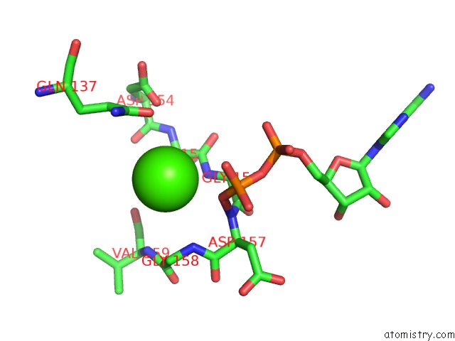
Mono view
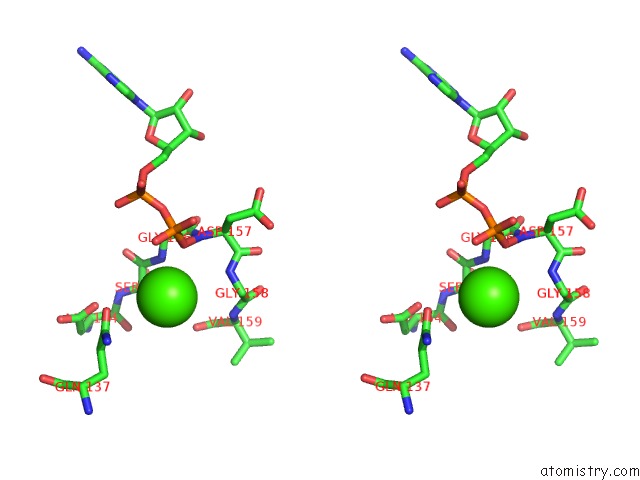
Stereo pair view
|
A full contact list of Calcium with other atoms in the Ca binding
site number 3 of Structure of the Actin-Tropomyosin-Myosin Complex (Rigor Atm 2) within 5.0Å range:
probe
|
atom
|
residue
|
distance (Å)
|
B
|
Occ
|
E:Ca377
b:12.4
occ:1.00
|
O3B
|
E:ADP376
|
3.3
|
10.0
|
1.0
|
N
|
E:GLY156
|
3.5
|
0.0
|
1.0
|
OE1
|
E:GLN137
|
3.8
|
0.0
|
1.0
|
C
|
E:SER155
|
4.0
|
0.0
|
1.0
|
N
|
E:GLY158
|
4.0
|
0.0
|
1.0
|
CA
|
E:GLY156
|
4.1
|
0.0
|
1.0
|
O1B
|
E:ADP376
|
4.1
|
11.2
|
1.0
|
CA
|
E:SER155
|
4.1
|
0.0
|
1.0
|
N
|
E:ASP157
|
4.2
|
0.0
|
1.0
|
PB
|
E:ADP376
|
4.3
|
10.8
|
1.0
|
CD
|
E:GLN137
|
4.4
|
0.0
|
1.0
|
CA
|
E:GLY158
|
4.5
|
0.0
|
1.0
|
C
|
E:GLY156
|
4.6
|
0.0
|
1.0
|
OD1
|
E:ASP154
|
4.7
|
0.0
|
1.0
|
O
|
E:VAL159
|
4.8
|
0.0
|
1.0
|
O
|
E:SER155
|
4.8
|
0.0
|
1.0
|
N
|
E:VAL159
|
4.9
|
0.0
|
1.0
|
CG
|
E:GLN137
|
5.0
|
0.0
|
1.0
|
N
|
E:SER155
|
5.0
|
0.0
|
1.0
|
NE2
|
E:GLN137
|
5.0
|
0.0
|
1.0
|
|
Calcium binding site 4 out
of 5 in 4a7h
Go back to
Calcium Binding Sites List in 4a7h
Calcium binding site 4 out
of 5 in the Structure of the Actin-Tropomyosin-Myosin Complex (Rigor Atm 2)
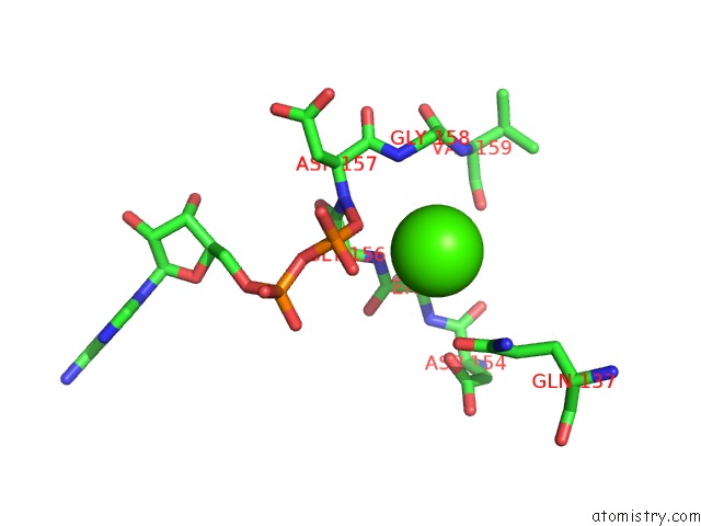
Mono view
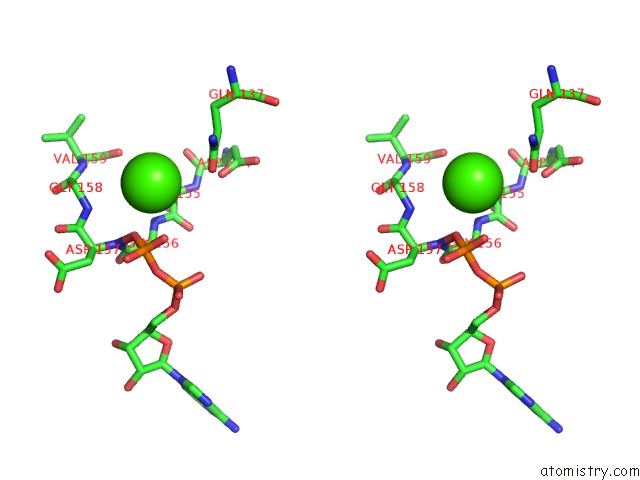
Stereo pair view
|
A full contact list of Calcium with other atoms in the Ca binding
site number 4 of Structure of the Actin-Tropomyosin-Myosin Complex (Rigor Atm 2) within 5.0Å range:
probe
|
atom
|
residue
|
distance (Å)
|
B
|
Occ
|
F:Ca377
b:12.4
occ:1.00
|
O3B
|
F:ADP376
|
3.3
|
10.0
|
1.0
|
N
|
F:GLY156
|
3.5
|
0.0
|
1.0
|
OE1
|
F:GLN137
|
3.8
|
0.0
|
1.0
|
C
|
F:SER155
|
4.0
|
0.0
|
1.0
|
N
|
F:GLY158
|
4.0
|
0.0
|
1.0
|
CA
|
F:GLY156
|
4.1
|
0.0
|
1.0
|
O1B
|
F:ADP376
|
4.1
|
11.2
|
1.0
|
CA
|
F:SER155
|
4.1
|
0.0
|
1.0
|
N
|
F:ASP157
|
4.2
|
0.0
|
1.0
|
PB
|
F:ADP376
|
4.3
|
10.8
|
1.0
|
CD
|
F:GLN137
|
4.4
|
0.0
|
1.0
|
CA
|
F:GLY158
|
4.5
|
0.0
|
1.0
|
C
|
F:GLY156
|
4.6
|
0.0
|
1.0
|
OD1
|
F:ASP154
|
4.7
|
0.0
|
1.0
|
O
|
F:VAL159
|
4.8
|
0.0
|
1.0
|
O
|
F:SER155
|
4.8
|
0.0
|
1.0
|
N
|
F:VAL159
|
4.9
|
0.0
|
1.0
|
CG
|
F:GLN137
|
5.0
|
0.0
|
1.0
|
N
|
F:SER155
|
5.0
|
0.0
|
1.0
|
NE2
|
F:GLN137
|
5.0
|
0.0
|
1.0
|
|
Calcium binding site 5 out
of 5 in 4a7h
Go back to
Calcium Binding Sites List in 4a7h
Calcium binding site 5 out
of 5 in the Structure of the Actin-Tropomyosin-Myosin Complex (Rigor Atm 2)
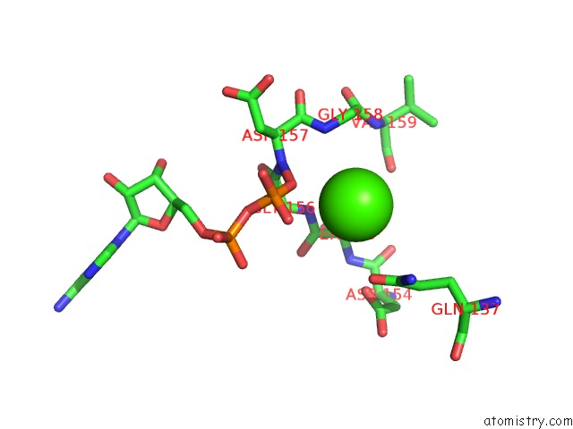
Mono view
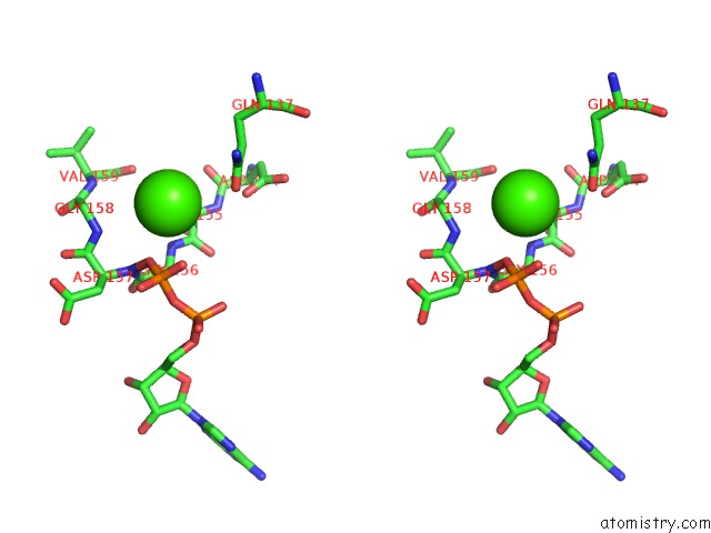
Stereo pair view
|
A full contact list of Calcium with other atoms in the Ca binding
site number 5 of Structure of the Actin-Tropomyosin-Myosin Complex (Rigor Atm 2) within 5.0Å range:
probe
|
atom
|
residue
|
distance (Å)
|
B
|
Occ
|
G:Ca377
b:12.4
occ:1.00
|
O3B
|
G:ADP376
|
3.3
|
10.0
|
1.0
|
N
|
G:GLY156
|
3.5
|
0.0
|
1.0
|
OE1
|
G:GLN137
|
3.8
|
0.0
|
1.0
|
C
|
G:SER155
|
4.0
|
0.0
|
1.0
|
N
|
G:GLY158
|
4.0
|
0.0
|
1.0
|
CA
|
G:GLY156
|
4.1
|
0.0
|
1.0
|
O1B
|
G:ADP376
|
4.1
|
11.2
|
1.0
|
CA
|
G:SER155
|
4.1
|
0.0
|
1.0
|
N
|
G:ASP157
|
4.2
|
0.0
|
1.0
|
PB
|
G:ADP376
|
4.3
|
10.8
|
1.0
|
CD
|
G:GLN137
|
4.4
|
0.0
|
1.0
|
CA
|
G:GLY158
|
4.5
|
0.0
|
1.0
|
C
|
G:GLY156
|
4.6
|
0.0
|
1.0
|
OD1
|
G:ASP154
|
4.7
|
0.0
|
1.0
|
O
|
G:VAL159
|
4.8
|
0.0
|
1.0
|
O
|
G:SER155
|
4.8
|
0.0
|
1.0
|
N
|
G:VAL159
|
4.9
|
0.0
|
1.0
|
CG
|
G:GLN137
|
5.0
|
0.0
|
1.0
|
N
|
G:SER155
|
5.0
|
0.0
|
1.0
|
NE2
|
G:GLN137
|
5.0
|
0.0
|
1.0
|
|
Reference:
E.Behrmann,
M.Muller,
P.A.Penczek,
H.G.Mannherz,
D.J.Manstein,
S.Raunser.
Structure of the Rigor Actin-Tropomyosin-Myosin Complex. Cell(Cambridge,Mass.) V. 150 327 2012.
ISSN: ISSN 0092-8674
PubMed: 22817895
DOI: 10.1016/J.CELL.2012.05.037
Page generated: Sat Jul 13 21:55:56 2024
|