Calcium »
PDB 4a42-4adj »
4a7l »
Calcium in PDB 4a7l: Structure of the Actin-Tropomyosin-Myosin Complex (Rigor Atm 1)
Enzymatic activity of Structure of the Actin-Tropomyosin-Myosin Complex (Rigor Atm 1)
All present enzymatic activity of Structure of the Actin-Tropomyosin-Myosin Complex (Rigor Atm 1):
3.6.4.1;
3.6.4.1;
Calcium Binding Sites:
The binding sites of Calcium atom in the Structure of the Actin-Tropomyosin-Myosin Complex (Rigor Atm 1)
(pdb code 4a7l). This binding sites where shown within
5.0 Angstroms radius around Calcium atom.
In total 5 binding sites of Calcium where determined in the Structure of the Actin-Tropomyosin-Myosin Complex (Rigor Atm 1), PDB code: 4a7l:
Jump to Calcium binding site number: 1; 2; 3; 4; 5;
In total 5 binding sites of Calcium where determined in the Structure of the Actin-Tropomyosin-Myosin Complex (Rigor Atm 1), PDB code: 4a7l:
Jump to Calcium binding site number: 1; 2; 3; 4; 5;
Calcium binding site 1 out of 5 in 4a7l
Go back to
Calcium binding site 1 out
of 5 in the Structure of the Actin-Tropomyosin-Myosin Complex (Rigor Atm 1)

Mono view
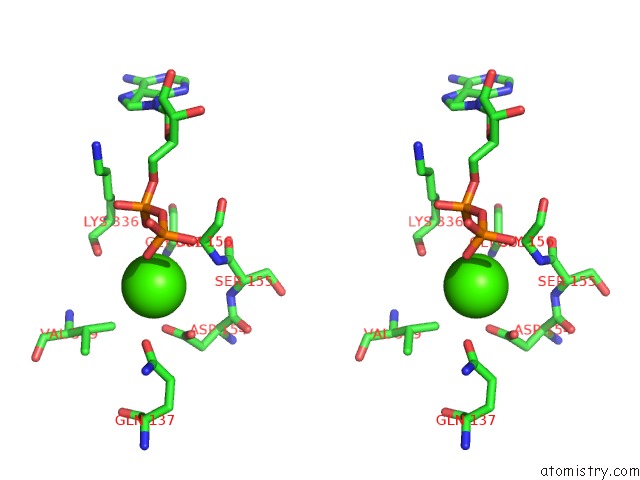
Stereo pair view
Mono view
Stereo pair view
A full contact list of Calcium with other atoms in the Ca binding
site number 1 of Structure of the Actin-Tropomyosin-Myosin Complex (Rigor Atm 1) within 5.0Å range:
|
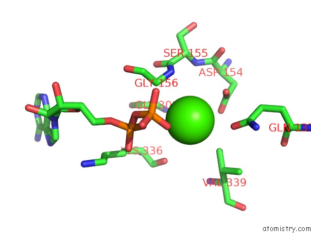
Calcium binding site 2 out of 5 in 4a7l
Go back to
Calcium binding site 2 out
of 5 in the Structure of the Actin-Tropomyosin-Myosin Complex (Rigor Atm 1)

Mono view

Stereo pair view
Mono view
Stereo pair view
A full contact list of Calcium with other atoms in the Ca binding
site number 2 of Structure of the Actin-Tropomyosin-Myosin Complex (Rigor Atm 1) within 5.0Å range:
|
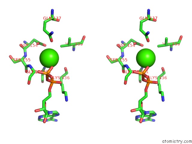
Calcium binding site 3 out of 5 in 4a7l
Go back to
Calcium binding site 3 out
of 5 in the Structure of the Actin-Tropomyosin-Myosin Complex (Rigor Atm 1)

Mono view

Stereo pair view
Mono view
Stereo pair view
A full contact list of Calcium with other atoms in the Ca binding
site number 3 of Structure of the Actin-Tropomyosin-Myosin Complex (Rigor Atm 1) within 5.0Å range:
|
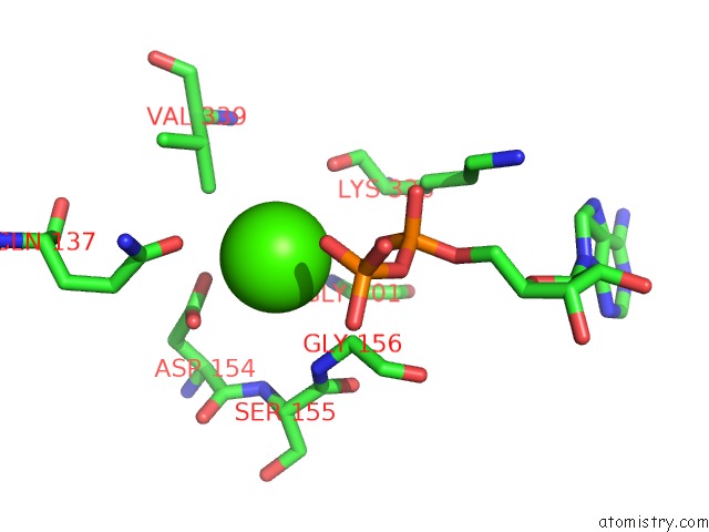
Calcium binding site 4 out of 5 in 4a7l
Go back to
Calcium binding site 4 out
of 5 in the Structure of the Actin-Tropomyosin-Myosin Complex (Rigor Atm 1)
Mono view
Stereo pair view
Mono view
Stereo pair view
A full contact list of Calcium with other atoms in the Ca binding
site number 4 of Structure of the Actin-Tropomyosin-Myosin Complex (Rigor Atm 1) within 5.0Å range:
|
Calcium binding site 5 out of 5 in 4a7l
Go back to
Calcium binding site 5 out
of 5 in the Structure of the Actin-Tropomyosin-Myosin Complex (Rigor Atm 1)
Mono view

Stereo pair view
Mono view
Stereo pair view
A full contact list of Calcium with other atoms in the Ca binding
site number 5 of Structure of the Actin-Tropomyosin-Myosin Complex (Rigor Atm 1) within 5.0Å range:
|
Reference:
E.Behrmann,
M.Muller,
P.A.Penczek,
H.G.Mannherz,
D.J.Manstein,
S.Raunser.
Structure of the Rigor Actin-Tropomyosin-Myosin Complex. Cell(Cambridge,Mass.) V. 150 327 2012.
ISSN: ISSN 0092-8674
PubMed: 22817895
DOI: 10.1016/J.CELL.2012.05.037
Page generated: Tue Jul 8 18:26:12 2025
ISSN: ISSN 0092-8674
PubMed: 22817895
DOI: 10.1016/J.CELL.2012.05.037
Last articles
Cl in 8AY7Cl in 8AY3
Cl in 8AY6
Cl in 8AXT
Cl in 8AXS
Cl in 8AXW
Cl in 8AXU
Cl in 8AXI
Cl in 8AXC
Cl in 8AXR