Calcium »
PDB 4a42-4adj »
4aag »
Calcium in PDB 4aag: Crystal Structure of the Mutant D75N I-Crei in Complex with Its Wild-Type Target in Presence of Ca at the Active Site (the Four Central Bases, 2NN Region, Are Composed By Gtac From 5' to 3')
Protein crystallography data
The structure of Crystal Structure of the Mutant D75N I-Crei in Complex with Its Wild-Type Target in Presence of Ca at the Active Site (the Four Central Bases, 2NN Region, Are Composed By Gtac From 5' to 3'), PDB code: 4aag
was solved by
R.Molina,
P.Redondo,
S.Stella,
M.Marenchino,
M.D'abramo,
F.Gervasio,
J.C.Epinat,
J.Valton,
S.Grizot,
P.Duchateau,
J.Prieto,
G.Montoya,
with X-Ray Crystallography technique. A brief refinement statistics is given in the table below:
| Resolution Low / High (Å) | 45.743 / 2.80 |
| Space group | P 2 21 21 |
| Cell size a, b, c (Å), α, β, γ (°) | 47.469, 71.413, 171.214, 90.00, 90.00, 90.00 |
| R / Rfree (%) | 17.35 / 24.97 |
Calcium Binding Sites:
The binding sites of Calcium atom in the Crystal Structure of the Mutant D75N I-Crei in Complex with Its Wild-Type Target in Presence of Ca at the Active Site (the Four Central Bases, 2NN Region, Are Composed By Gtac From 5' to 3')
(pdb code 4aag). This binding sites where shown within
5.0 Angstroms radius around Calcium atom.
In total 2 binding sites of Calcium where determined in the Crystal Structure of the Mutant D75N I-Crei in Complex with Its Wild-Type Target in Presence of Ca at the Active Site (the Four Central Bases, 2NN Region, Are Composed By Gtac From 5' to 3'), PDB code: 4aag:
Jump to Calcium binding site number: 1; 2;
Calcium binding site 1 out of 2 in 4aag
Go back to
Calcium binding site 1 out
of 2 in the Crystal Structure of the Mutant D75N I-Crei in Complex with Its Wild-Type Target in Presence of Ca at the Active Site (the Four Central Bases, 2NN Region, Are Composed By Gtac From 5' to 3')
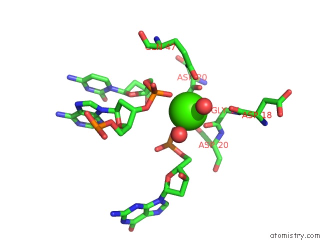
Mono view
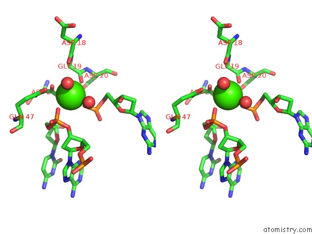
Stereo pair view
Mono view
Stereo pair view
A full contact list of Calcium with other atoms in the Ca binding
site number 1 of Crystal Structure of the Mutant D75N I-Crei in Complex with Its Wild-Type Target in Presence of Ca at the Active Site (the Four Central Bases, 2NN Region, Are Composed By Gtac From 5' to 3') within 5.0Å range:
|
Calcium binding site 2 out of 2 in 4aag
Go back to
Calcium binding site 2 out
of 2 in the Crystal Structure of the Mutant D75N I-Crei in Complex with Its Wild-Type Target in Presence of Ca at the Active Site (the Four Central Bases, 2NN Region, Are Composed By Gtac From 5' to 3')
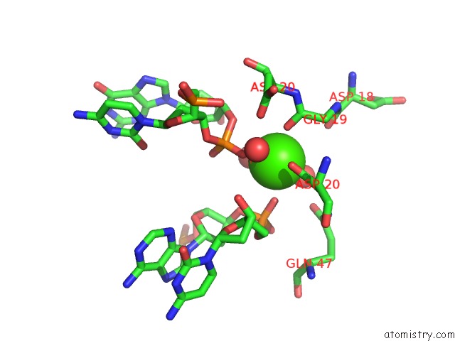
Mono view
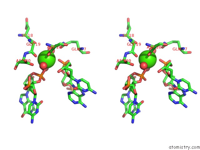
Stereo pair view
Mono view
Stereo pair view
A full contact list of Calcium with other atoms in the Ca binding
site number 2 of Crystal Structure of the Mutant D75N I-Crei in Complex with Its Wild-Type Target in Presence of Ca at the Active Site (the Four Central Bases, 2NN Region, Are Composed By Gtac From 5' to 3') within 5.0Å range:
|
Reference:
R.Molina,
P.Redondo,
S.Stella,
M.Marenchino,
M.D'abramo,
F.L.Gervasio,
J.Charles Epinat,
J.Valton,
S.Grizot,
P.Duchateau,
J.Prieto,
G.Montoya.
Non-Specific Protein-Dna Interactions Control I-Crei Target Binding and Cleavage. Nucleic Acids Res. V. 40 6936 2012.
ISSN: ISSN 0305-1048
PubMed: 22495931
DOI: 10.1093/NAR/GKS320
Page generated: Tue Jul 8 18:27:17 2025
ISSN: ISSN 0305-1048
PubMed: 22495931
DOI: 10.1093/NAR/GKS320
Last articles
Fe in 2YXOFe in 2YRS
Fe in 2YXC
Fe in 2YNM
Fe in 2YVJ
Fe in 2YP1
Fe in 2YU2
Fe in 2YU1
Fe in 2YQB
Fe in 2YOO