Calcium »
PDB 4ae2-4aqe »
4al9 »
Calcium in PDB 4al9: Crystal Structure of the Lectin Pa-Il From Pseudomonas Aeruginoas in Complex with Melibiose
Protein crystallography data
The structure of Crystal Structure of the Lectin Pa-Il From Pseudomonas Aeruginoas in Complex with Melibiose, PDB code: 4al9
was solved by
B.Blanchard,
A.Imberty,
A.Varrot,
with X-Ray Crystallography technique. A brief refinement statistics is given in the table below:
| Resolution Low / High (Å) | 30.95 / 1.75 |
| Space group | P 1 |
| Cell size a, b, c (Å), α, β, γ (°) | 50.070, 58.130, 75.940, 101.13, 92.89, 100.98 |
| R / Rfree (%) | 16.659 / 21.134 |
Calcium Binding Sites:
The binding sites of Calcium atom in the Crystal Structure of the Lectin Pa-Il From Pseudomonas Aeruginoas in Complex with Melibiose
(pdb code 4al9). This binding sites where shown within
5.0 Angstroms radius around Calcium atom.
In total 8 binding sites of Calcium where determined in the Crystal Structure of the Lectin Pa-Il From Pseudomonas Aeruginoas in Complex with Melibiose, PDB code: 4al9:
Jump to Calcium binding site number: 1; 2; 3; 4; 5; 6; 7; 8;
In total 8 binding sites of Calcium where determined in the Crystal Structure of the Lectin Pa-Il From Pseudomonas Aeruginoas in Complex with Melibiose, PDB code: 4al9:
Jump to Calcium binding site number: 1; 2; 3; 4; 5; 6; 7; 8;
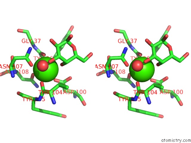
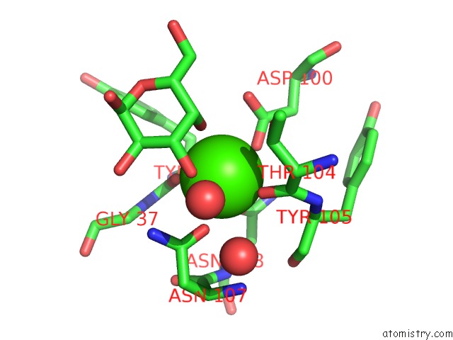
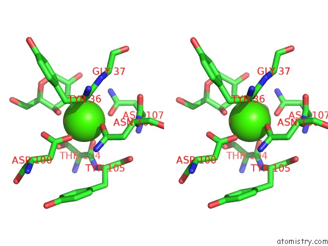
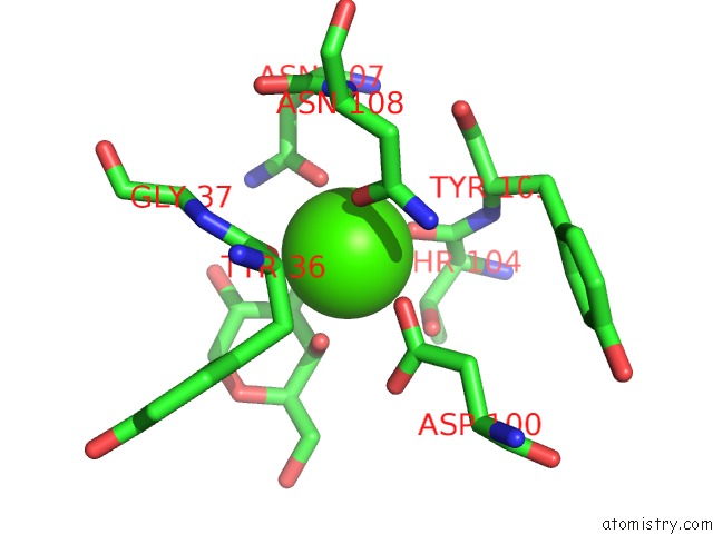
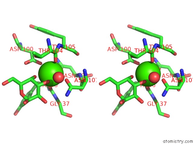
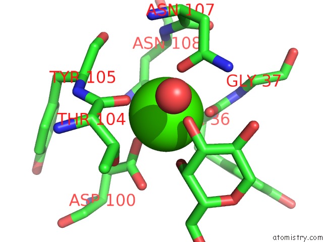
Calcium binding site 1 out of 8 in 4al9
Go back to
Calcium binding site 1 out
of 8 in the Crystal Structure of the Lectin Pa-Il From Pseudomonas Aeruginoas in Complex with Melibiose
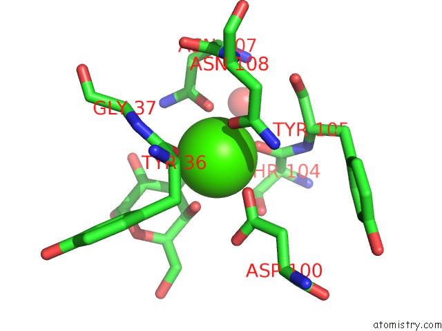
Mono view
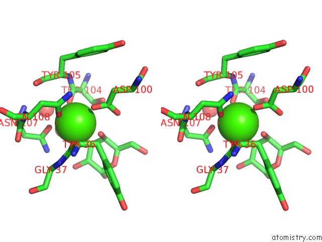
Stereo pair view
Mono view
Stereo pair view
A full contact list of Calcium with other atoms in the Ca binding
site number 1 of Crystal Structure of the Lectin Pa-Il From Pseudomonas Aeruginoas in Complex with Melibiose within 5.0Å range:
|
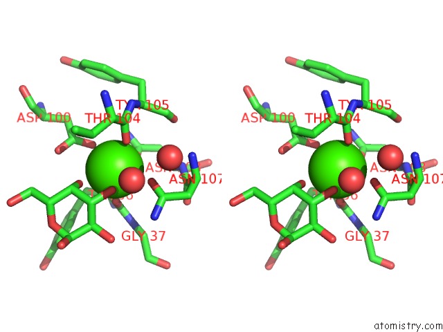
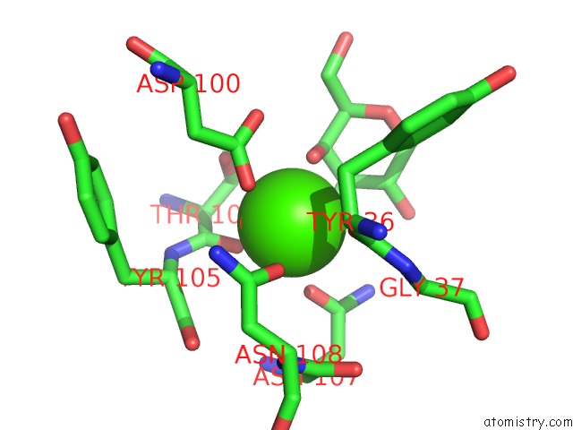
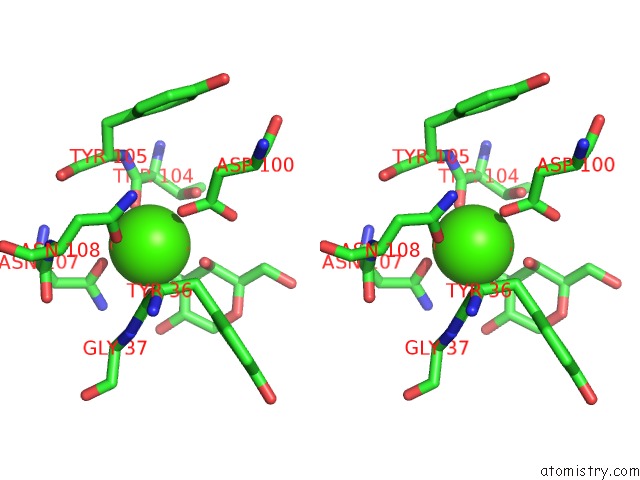
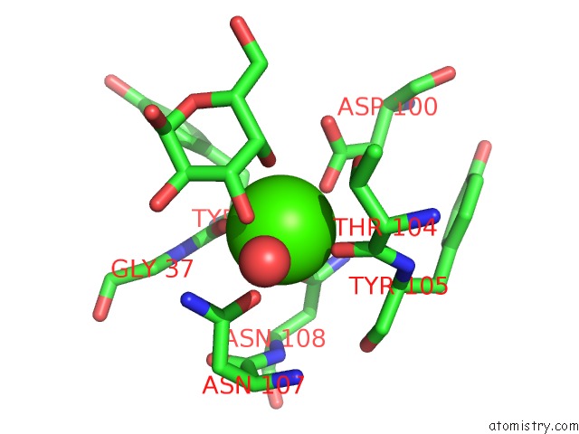
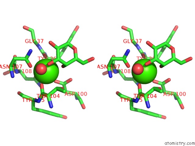
Calcium binding site 2 out of 8 in 4al9
Go back to
Calcium binding site 2 out
of 8 in the Crystal Structure of the Lectin Pa-Il From Pseudomonas Aeruginoas in Complex with Melibiose
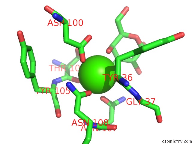
Mono view
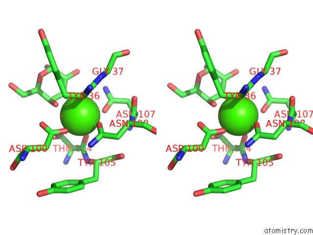
Stereo pair view
Mono view
Stereo pair view
A full contact list of Calcium with other atoms in the Ca binding
site number 2 of Crystal Structure of the Lectin Pa-Il From Pseudomonas Aeruginoas in Complex with Melibiose within 5.0Å range:
|
Calcium binding site 3 out of 8 in 4al9
Go back to
Calcium binding site 3 out
of 8 in the Crystal Structure of the Lectin Pa-Il From Pseudomonas Aeruginoas in Complex with Melibiose

Mono view
Stereo pair view
Mono view
Stereo pair view
A full contact list of Calcium with other atoms in the Ca binding
site number 3 of Crystal Structure of the Lectin Pa-Il From Pseudomonas Aeruginoas in Complex with Melibiose within 5.0Å range:
|
Calcium binding site 4 out of 8 in 4al9
Go back to
Calcium binding site 4 out
of 8 in the Crystal Structure of the Lectin Pa-Il From Pseudomonas Aeruginoas in Complex with Melibiose
Mono view
Stereo pair view
Mono view
Stereo pair view
A full contact list of Calcium with other atoms in the Ca binding
site number 4 of Crystal Structure of the Lectin Pa-Il From Pseudomonas Aeruginoas in Complex with Melibiose within 5.0Å range:
|
Calcium binding site 5 out of 8 in 4al9
Go back to
Calcium binding site 5 out
of 8 in the Crystal Structure of the Lectin Pa-Il From Pseudomonas Aeruginoas in Complex with Melibiose
Mono view
Stereo pair view
Mono view
Stereo pair view
A full contact list of Calcium with other atoms in the Ca binding
site number 5 of Crystal Structure of the Lectin Pa-Il From Pseudomonas Aeruginoas in Complex with Melibiose within 5.0Å range:
|
Calcium binding site 6 out of 8 in 4al9
Go back to
Calcium binding site 6 out
of 8 in the Crystal Structure of the Lectin Pa-Il From Pseudomonas Aeruginoas in Complex with Melibiose
Mono view
Stereo pair view
Mono view
Stereo pair view
A full contact list of Calcium with other atoms in the Ca binding
site number 6 of Crystal Structure of the Lectin Pa-Il From Pseudomonas Aeruginoas in Complex with Melibiose within 5.0Å range:
|
Calcium binding site 7 out of 8 in 4al9
Go back to
Calcium binding site 7 out
of 8 in the Crystal Structure of the Lectin Pa-Il From Pseudomonas Aeruginoas in Complex with Melibiose
Mono view
Stereo pair view
Mono view
Stereo pair view
A full contact list of Calcium with other atoms in the Ca binding
site number 7 of Crystal Structure of the Lectin Pa-Il From Pseudomonas Aeruginoas in Complex with Melibiose within 5.0Å range:
|
Calcium binding site 8 out of 8 in 4al9
Go back to
Calcium binding site 8 out
of 8 in the Crystal Structure of the Lectin Pa-Il From Pseudomonas Aeruginoas in Complex with Melibiose
Mono view
Stereo pair view
Mono view
Stereo pair view
A full contact list of Calcium with other atoms in the Ca binding
site number 8 of Crystal Structure of the Lectin Pa-Il From Pseudomonas Aeruginoas in Complex with Melibiose within 5.0Å range:
|
Reference:
B.Blanchard,
A.Imberty,
A.Varrot.
Secondary Sugar Binding Site Identified For Leca Lectin From Pseudomonas Aeruginosa. Proteins V. 82 1060 2014.
ISSN: ISSN 0887-3585
PubMed: 24123124
DOI: 10.1002/PROT.24430
Page generated: Sat Jul 13 22:05:41 2024
ISSN: ISSN 0887-3585
PubMed: 24123124
DOI: 10.1002/PROT.24430
Last articles
Zn in 9J0NZn in 9J0O
Zn in 9J0P
Zn in 9FJX
Zn in 9EKB
Zn in 9C0F
Zn in 9CAH
Zn in 9CH0
Zn in 9CH3
Zn in 9CH1