Calcium »
PDB 4aqi-4ayp »
4aqj »
Calcium in PDB 4aqj: Structure of Human S100A7 D24G Bound to Zinc and Calcium
Protein crystallography data
The structure of Structure of Human S100A7 D24G Bound to Zinc and Calcium, PDB code: 4aqj
was solved by
J.I.Murray,
M.L.Tonkin,
A.L.Whiting,
F.Peng,
B.Farnell,
F.Hof,
M.J.Boulanger,
with X-Ray Crystallography technique. A brief refinement statistics is given in the table below:
| Resolution Low / High (Å) | 34.76 / 1.60 |
| Space group | P 43 21 2 |
| Cell size a, b, c (Å), α, β, γ (°) | 51.480, 51.480, 117.230, 90.00, 90.00, 90.00 |
| R / Rfree (%) | 18.901 / 22.532 |
Other elements in 4aqj:
The structure of Structure of Human S100A7 D24G Bound to Zinc and Calcium also contains other interesting chemical elements:
| Chlorine | (Cl) | 1 atom |
| Zinc | (Zn) | 1 atom |
Calcium Binding Sites:
The binding sites of Calcium atom in the Structure of Human S100A7 D24G Bound to Zinc and Calcium
(pdb code 4aqj). This binding sites where shown within
5.0 Angstroms radius around Calcium atom.
In total only one binding site of Calcium was determined in the Structure of Human S100A7 D24G Bound to Zinc and Calcium, PDB code: 4aqj:
In total only one binding site of Calcium was determined in the Structure of Human S100A7 D24G Bound to Zinc and Calcium, PDB code: 4aqj:
Calcium binding site 1 out of 1 in 4aqj
Go back to
Calcium binding site 1 out
of 1 in the Structure of Human S100A7 D24G Bound to Zinc and Calcium
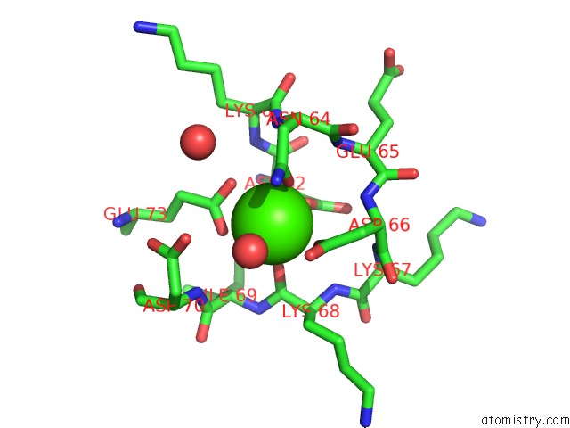
Mono view
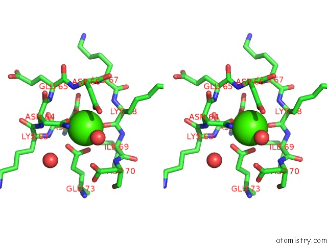
Stereo pair view
Mono view
Stereo pair view
A full contact list of Calcium with other atoms in the Ca binding
site number 1 of Structure of Human S100A7 D24G Bound to Zinc and Calcium within 5.0Å range:
|
Reference:
J.I.Murray,
M.L.Tonkin,
A.L.Whiting,
F.Peng,
B.Farnell,
J.T.Cullen,
F.Hof,
M.J.Boulanger.
Structural Characterization of S100A15 Reveals A Novel Zinc Coordination Site Among S100 Proteins and Altered Surface Chemistry with Functional Implications For Receptor Binding. Bmc Struct.Biol. V. 12 16 2012.
ISSN: ISSN 1472-6807
PubMed: 22747601
DOI: 10.1186/1472-6807-12-16
Page generated: Tue Jul 8 18:39:20 2025
ISSN: ISSN 1472-6807
PubMed: 22747601
DOI: 10.1186/1472-6807-12-16
Last articles
F in 7OP1F in 7ORF
F in 7OP8
F in 7ORE
F in 7OP5
F in 7OL8
F in 7OL7
F in 7OL3
F in 7OOB
F in 7OHN