Calcium »
PDB 4aqi-4ayp »
4ate »
Calcium in PDB 4ate: High Resolution Crystal Structure of Beta-Porphyranase A From Zobellia Galactanivorans
Enzymatic activity of High Resolution Crystal Structure of Beta-Porphyranase A From Zobellia Galactanivorans
All present enzymatic activity of High Resolution Crystal Structure of Beta-Porphyranase A From Zobellia Galactanivorans:
3.2.1.178;
3.2.1.178;
Protein crystallography data
The structure of High Resolution Crystal Structure of Beta-Porphyranase A From Zobellia Galactanivorans, PDB code: 4ate
was solved by
J.H.Hehemann,
G.Correc,
M.Jam,
G.Michel,
M.Czjzek,
with X-Ray Crystallography technique. A brief refinement statistics is given in the table below:
| Resolution Low / High (Å) | 25.86 / 1.10 |
| Space group | P 31 2 1 |
| Cell size a, b, c (Å), α, β, γ (°) | 70.270, 70.270, 92.470, 90.00, 90.00, 120.00 |
| R / Rfree (%) | 12.4 / 15.2 |
Other elements in 4ate:
The structure of High Resolution Crystal Structure of Beta-Porphyranase A From Zobellia Galactanivorans also contains other interesting chemical elements:
| Chlorine | (Cl) | 1 atom |
Calcium Binding Sites:
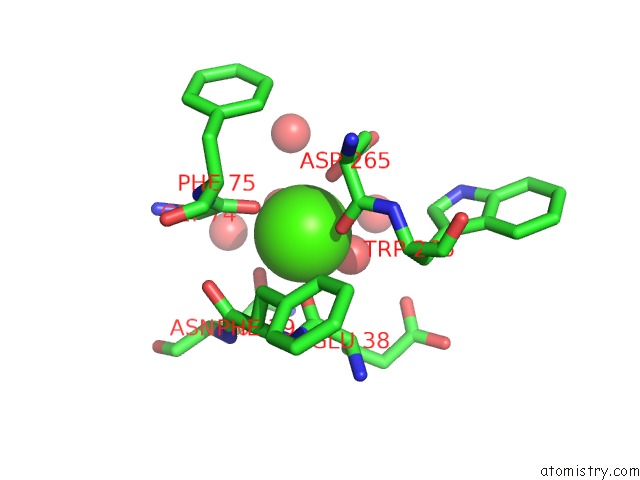
The binding sites of Calcium atom in the High Resolution Crystal Structure of Beta-Porphyranase A From Zobellia Galactanivorans
(pdb code 4ate). This binding sites where shown within
5.0 Angstroms radius around Calcium atom.
In total only one binding site of Calcium was determined in the High Resolution Crystal Structure of Beta-Porphyranase A From Zobellia Galactanivorans, PDB code: 4ate:
In total only one binding site of Calcium was determined in the High Resolution Crystal Structure of Beta-Porphyranase A From Zobellia Galactanivorans, PDB code: 4ate:
Calcium binding site 1 out of 1 in 4ate
Go back to
Calcium binding site 1 out
of 1 in the High Resolution Crystal Structure of Beta-Porphyranase A From Zobellia Galactanivorans
Mono view

Stereo pair view
Mono view
Stereo pair view
A full contact list of Calcium with other atoms in the Ca binding
site number 1 of High Resolution Crystal Structure of Beta-Porphyranase A From Zobellia Galactanivorans within 5.0Å range:
|
Reference:
J.H.Hehemann,
G.Correc,
F.Thomas,
T.Bernard,
T.Barbeyron,
M.Jam,
W.Helbert,
G.Michel,
M.Czjzek.
Biochemical and Structural Characterization of the Complex Agarolytic Enzyme System From the Marine Bacterium Zobellia Galactanivorans. J.Biol.Chem. V. 287 30571 2012.
ISSN: ISSN 0021-9258
PubMed: 22778272
DOI: 10.1074/JBC.M112.377184
Page generated: Tue Jul 8 18:40:34 2025
ISSN: ISSN 0021-9258
PubMed: 22778272
DOI: 10.1074/JBC.M112.377184
Last articles
Cl in 5I60Cl in 5I5W
Cl in 5I5T
Cl in 5I5S
Cl in 5I5V
Cl in 5I5U
Cl in 5I58
Cl in 5I54
Cl in 5I3T
Cl in 5I56