Calcium »
PDB 4aqi-4ayp »
4avt »
Calcium in PDB 4avt: Structure of Cphpc Bound to Serum Amyloid P Component
Protein crystallography data
The structure of Structure of Cphpc Bound to Serum Amyloid P Component, PDB code: 4avt
was solved by
S.E.Kolstoe,
A.Purvis,
S.P.Wood,
with X-Ray Crystallography technique. A brief refinement statistics is given in the table below:
| Resolution Low / High (Å) | 24.942 / 3.20 |
| Space group | P 43 21 2 |
| Cell size a, b, c (Å), α, β, γ (°) | 230.864, 230.864, 281.393, 90.00, 90.00, 90.00 |
| R / Rfree (%) | 18.89 / 19.74 |
Calcium Binding Sites:
Pages:
>>> Page 1 <<< Page 2, Binding sites: 11 - 20;Binding sites:
The binding sites of Calcium atom in the Structure of Cphpc Bound to Serum Amyloid P Component (pdb code 4avt). This binding sites where shown within 5.0 Angstroms radius around Calcium atom.In total 20 binding sites of Calcium where determined in the Structure of Cphpc Bound to Serum Amyloid P Component, PDB code: 4avt:
Jump to Calcium binding site number: 1; 2; 3; 4; 5; 6; 7; 8; 9; 10;
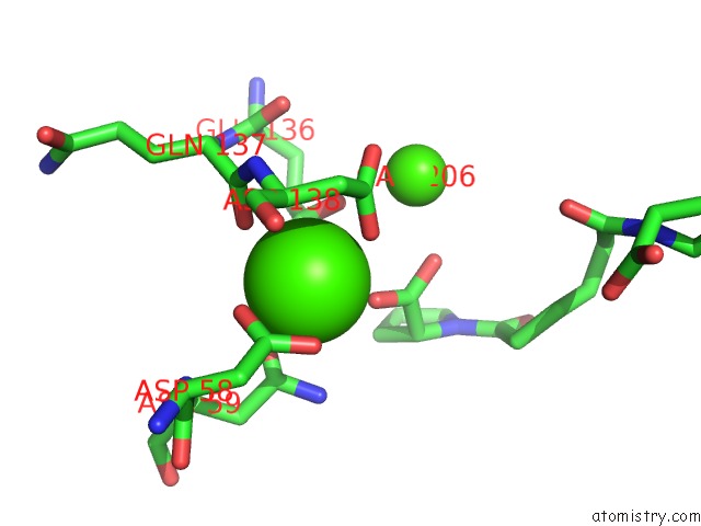
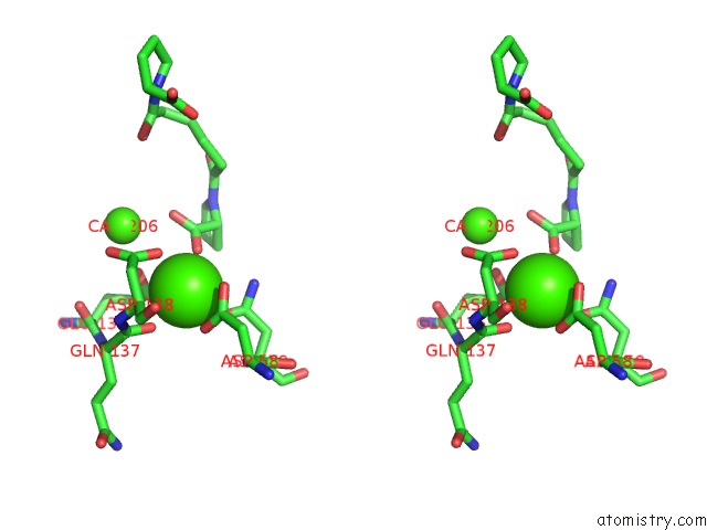
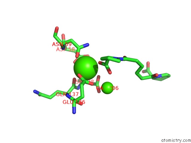
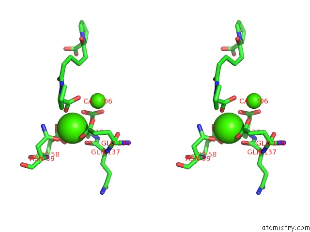
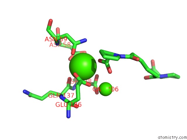
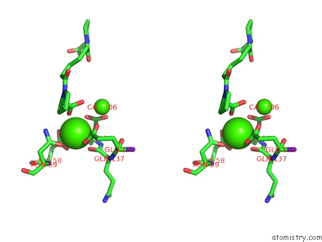
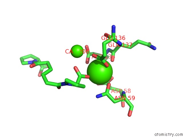
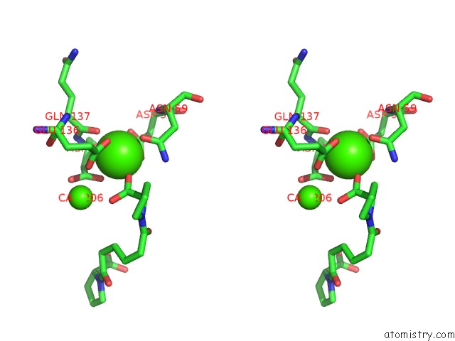
Calcium binding site 1 out of 20 in 4avt
Go back to
Calcium binding site 1 out
of 20 in the Structure of Cphpc Bound to Serum Amyloid P Component
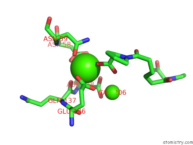
Mono view
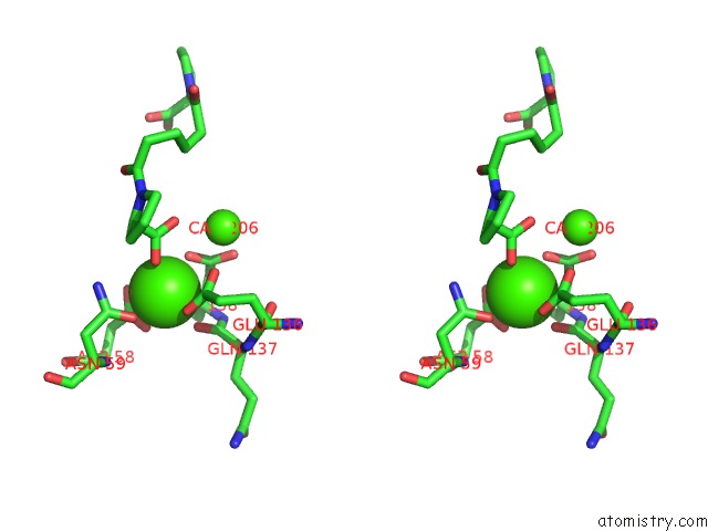
Stereo pair view
Mono view
Stereo pair view
A full contact list of Calcium with other atoms in the Ca binding
site number 1 of Structure of Cphpc Bound to Serum Amyloid P Component within 5.0Å range:
|
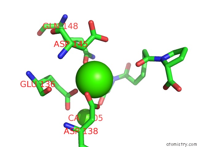
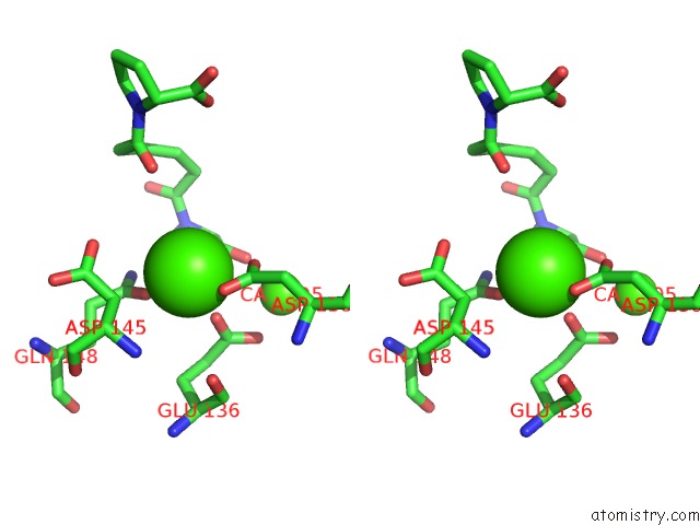
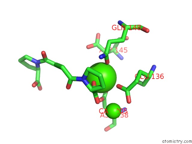
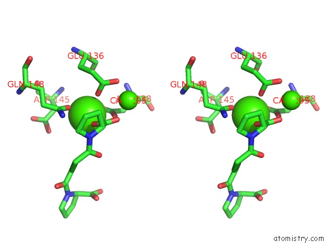
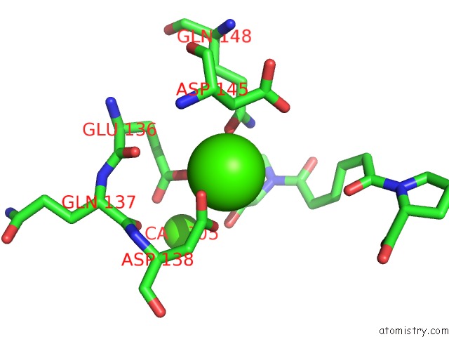
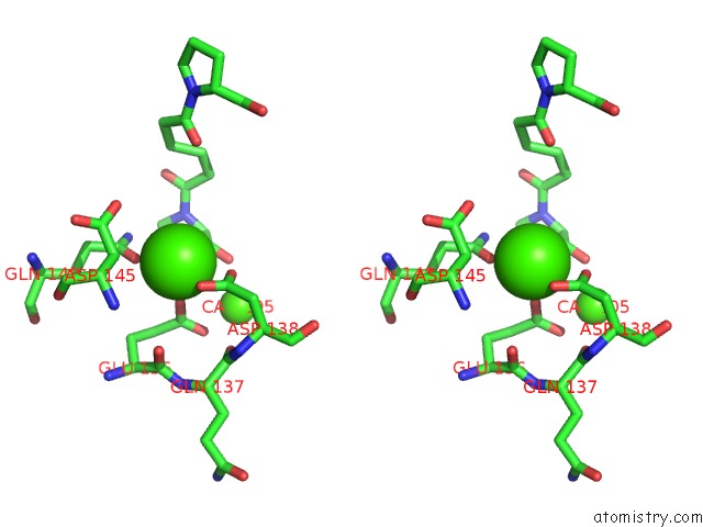
Calcium binding site 2 out of 20 in 4avt
Go back to
Calcium binding site 2 out
of 20 in the Structure of Cphpc Bound to Serum Amyloid P Component
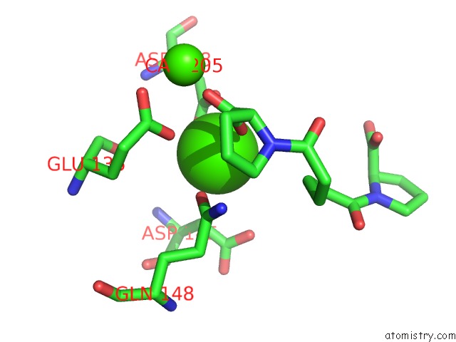
Mono view
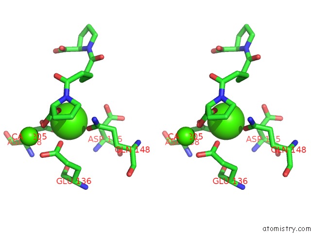
Stereo pair view
Mono view
Stereo pair view
A full contact list of Calcium with other atoms in the Ca binding
site number 2 of Structure of Cphpc Bound to Serum Amyloid P Component within 5.0Å range:
|
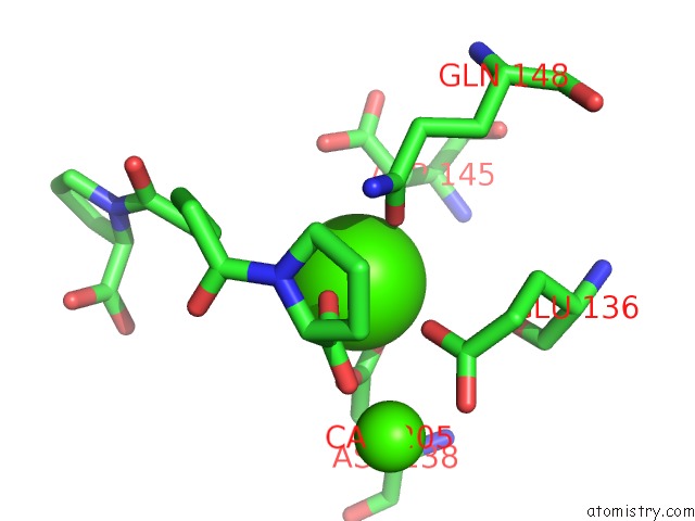
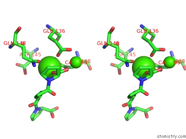
Calcium binding site 3 out of 20 in 4avt
Go back to
Calcium binding site 3 out
of 20 in the Structure of Cphpc Bound to Serum Amyloid P Component
Mono view
Stereo pair view
Mono view
Stereo pair view
A full contact list of Calcium with other atoms in the Ca binding
site number 3 of Structure of Cphpc Bound to Serum Amyloid P Component within 5.0Å range:
|
Calcium binding site 4 out of 20 in 4avt
Go back to
Calcium binding site 4 out
of 20 in the Structure of Cphpc Bound to Serum Amyloid P Component
Mono view
Stereo pair view
Mono view
Stereo pair view
A full contact list of Calcium with other atoms in the Ca binding
site number 4 of Structure of Cphpc Bound to Serum Amyloid P Component within 5.0Å range:
|
Calcium binding site 5 out of 20 in 4avt
Go back to
Calcium binding site 5 out
of 20 in the Structure of Cphpc Bound to Serum Amyloid P Component
Mono view
Stereo pair view
Mono view
Stereo pair view
A full contact list of Calcium with other atoms in the Ca binding
site number 5 of Structure of Cphpc Bound to Serum Amyloid P Component within 5.0Å range:
|
Calcium binding site 6 out of 20 in 4avt
Go back to
Calcium binding site 6 out
of 20 in the Structure of Cphpc Bound to Serum Amyloid P Component
Mono view
Stereo pair view
Mono view
Stereo pair view
A full contact list of Calcium with other atoms in the Ca binding
site number 6 of Structure of Cphpc Bound to Serum Amyloid P Component within 5.0Å range:
|
Calcium binding site 7 out of 20 in 4avt
Go back to
Calcium binding site 7 out
of 20 in the Structure of Cphpc Bound to Serum Amyloid P Component
Mono view
Stereo pair view
Mono view
Stereo pair view
A full contact list of Calcium with other atoms in the Ca binding
site number 7 of Structure of Cphpc Bound to Serum Amyloid P Component within 5.0Å range:
|
Calcium binding site 8 out of 20 in 4avt
Go back to
Calcium binding site 8 out
of 20 in the Structure of Cphpc Bound to Serum Amyloid P Component
Mono view
Stereo pair view
Mono view
Stereo pair view
A full contact list of Calcium with other atoms in the Ca binding
site number 8 of Structure of Cphpc Bound to Serum Amyloid P Component within 5.0Å range:
|
Calcium binding site 9 out of 20 in 4avt
Go back to
Calcium binding site 9 out
of 20 in the Structure of Cphpc Bound to Serum Amyloid P Component
Mono view
Stereo pair view
Mono view
Stereo pair view
A full contact list of Calcium with other atoms in the Ca binding
site number 9 of Structure of Cphpc Bound to Serum Amyloid P Component within 5.0Å range:
|
Calcium binding site 10 out of 20 in 4avt
Go back to
Calcium binding site 10 out
of 20 in the Structure of Cphpc Bound to Serum Amyloid P Component
Mono view
Stereo pair view
Mono view
Stereo pair view
A full contact list of Calcium with other atoms in the Ca binding
site number 10 of Structure of Cphpc Bound to Serum Amyloid P Component within 5.0Å range:
|
Reference:
S.E.Kolstoe,
M.C.Jenvey,
A.Purvis,
M.E.Light,
D.Thompson,
P.Hughes,
M.B.Pepys,
S.P.Wood.
Interaction of Serum Amyloid P Component with Hexanoyl Bis(D-Proline) (Cphpc) Acta Crystallogr.,Sect.D V. 70 2232 2014.
ISSN: ISSN 0907-4449
PubMed: 25084341
DOI: 10.1107/S1399004714013455
Page generated: Sat Jul 13 22:21:13 2024
ISSN: ISSN 0907-4449
PubMed: 25084341
DOI: 10.1107/S1399004714013455
Last articles
Zn in 9J0NZn in 9J0O
Zn in 9J0P
Zn in 9FJX
Zn in 9EKB
Zn in 9C0F
Zn in 9CAH
Zn in 9CH0
Zn in 9CH3
Zn in 9CH1