Calcium »
PDB 4ayq-4bae »
4b3v »
Calcium in PDB 4b3v: Crystal Structure of the Rubella Virus Glycoprotein E1 in Its Post- Fusion Form Crystallized in Presence of 20MM of Calcium Acetate
Protein crystallography data
The structure of Crystal Structure of the Rubella Virus Glycoprotein E1 in Its Post- Fusion Form Crystallized in Presence of 20MM of Calcium Acetate, PDB code: 4b3v
was solved by
M.C.Vaney,
R.M.Dubois,
M.A.Tortorici,
F.A.Rey,
with X-Ray Crystallography technique. A brief refinement statistics is given in the table below:
| Resolution Low / High (Å) | 28.99 / 1.98 |
| Space group | P 21 21 21 |
| Cell size a, b, c (Å), α, β, γ (°) | 121.272, 126.163, 129.375, 90.00, 90.00, 90.00 |
| R / Rfree (%) | 18.5 / 19.8 |
Other elements in 4b3v:
The structure of Crystal Structure of the Rubella Virus Glycoprotein E1 in Its Post- Fusion Form Crystallized in Presence of 20MM of Calcium Acetate also contains other interesting chemical elements:
| Sodium | (Na) | 1 atom |
Calcium Binding Sites:
The binding sites of Calcium atom in the Crystal Structure of the Rubella Virus Glycoprotein E1 in Its Post- Fusion Form Crystallized in Presence of 20MM of Calcium Acetate
(pdb code 4b3v). This binding sites where shown within
5.0 Angstroms radius around Calcium atom.
In total 3 binding sites of Calcium where determined in the Crystal Structure of the Rubella Virus Glycoprotein E1 in Its Post- Fusion Form Crystallized in Presence of 20MM of Calcium Acetate, PDB code: 4b3v:
Jump to Calcium binding site number: 1; 2; 3;
In total 3 binding sites of Calcium where determined in the Crystal Structure of the Rubella Virus Glycoprotein E1 in Its Post- Fusion Form Crystallized in Presence of 20MM of Calcium Acetate, PDB code: 4b3v:
Jump to Calcium binding site number: 1; 2; 3;
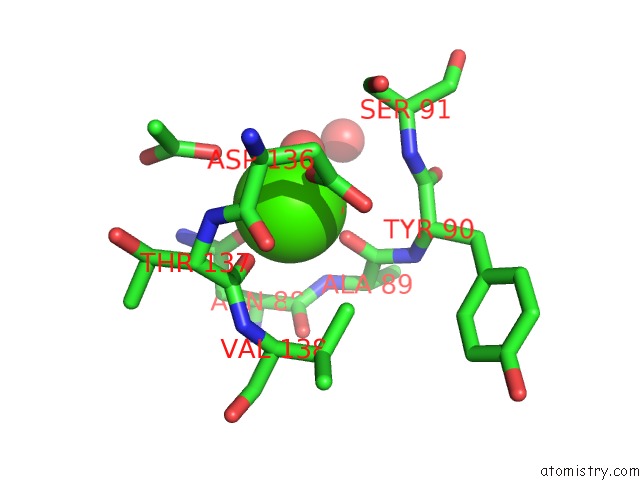
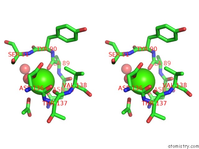
Calcium binding site 1 out of 3 in 4b3v
Go back to
Calcium binding site 1 out
of 3 in the Crystal Structure of the Rubella Virus Glycoprotein E1 in Its Post- Fusion Form Crystallized in Presence of 20MM of Calcium Acetate
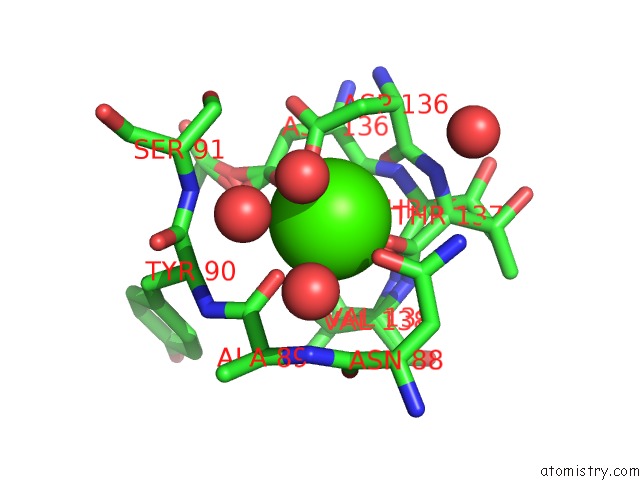
Mono view
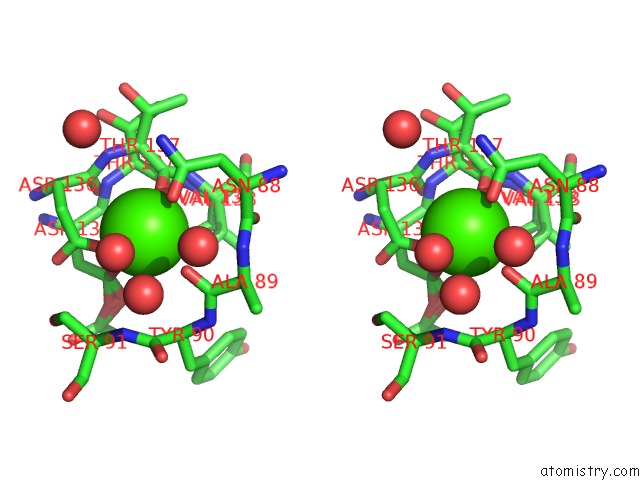
Stereo pair view
Mono view
Stereo pair view
A full contact list of Calcium with other atoms in the Ca binding
site number 1 of Crystal Structure of the Rubella Virus Glycoprotein E1 in Its Post- Fusion Form Crystallized in Presence of 20MM of Calcium Acetate within 5.0Å range:
|
Calcium binding site 2 out of 3 in 4b3v
Go back to
Calcium binding site 2 out
of 3 in the Crystal Structure of the Rubella Virus Glycoprotein E1 in Its Post- Fusion Form Crystallized in Presence of 20MM of Calcium Acetate
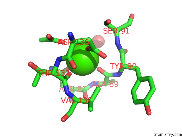
Mono view
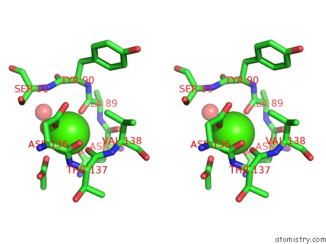
Stereo pair view
Mono view
Stereo pair view
A full contact list of Calcium with other atoms in the Ca binding
site number 2 of Crystal Structure of the Rubella Virus Glycoprotein E1 in Its Post- Fusion Form Crystallized in Presence of 20MM of Calcium Acetate within 5.0Å range:
|
Calcium binding site 3 out of 3 in 4b3v
Go back to
Calcium binding site 3 out
of 3 in the Crystal Structure of the Rubella Virus Glycoprotein E1 in Its Post- Fusion Form Crystallized in Presence of 20MM of Calcium Acetate
Mono view
Stereo pair view
Mono view
Stereo pair view
A full contact list of Calcium with other atoms in the Ca binding
site number 3 of Crystal Structure of the Rubella Virus Glycoprotein E1 in Its Post- Fusion Form Crystallized in Presence of 20MM of Calcium Acetate within 5.0Å range:
|
Reference:
R.M.Dubois,
M.C.Vaney,
M.A.Tortorici,
R.A.Kurdi,
G.Barba-Spaeth,
T.Krey,
F.A.Rey.
Functional and Evolutionary Insight From the Crystal Structure of Rubella Virus Protein E1. Nature V. 493 552 2013.
ISSN: ISSN 0028-0836
PubMed: 23292515
DOI: 10.1038/NATURE11741
Page generated: Tue Jul 8 18:50:15 2025
ISSN: ISSN 0028-0836
PubMed: 23292515
DOI: 10.1038/NATURE11741
Last articles
Cl in 5H4ZCl in 5H3G
Cl in 5GZX
Cl in 5H22
Cl in 5GY0
Cl in 5H0Z
Cl in 5H0Y
Cl in 5GZK
Cl in 5GXZ
Cl in 5GWF