Calcium »
PDB 4ayq-4bae »
4b9b »
Calcium in PDB 4b9b: The Structure of the Omega Aminotransferase From Pseudomonas Aeruginosa
Enzymatic activity of The Structure of the Omega Aminotransferase From Pseudomonas Aeruginosa
All present enzymatic activity of The Structure of the Omega Aminotransferase From Pseudomonas Aeruginosa:
2.6.1.18;
2.6.1.18;
Protein crystallography data
The structure of The Structure of the Omega Aminotransferase From Pseudomonas Aeruginosa, PDB code: 4b9b
was solved by
C.Sayer,
M.N.Isupov,
A.Westlake,
J.A.Littlechild,
with X-Ray Crystallography technique. A brief refinement statistics is given in the table below:
| Resolution Low / High (Å) | 66.58 / 1.64 |
| Space group | P 1 21 1 |
| Cell size a, b, c (Å), α, β, γ (°) | 80.380, 133.160, 161.960, 90.00, 91.75, 90.00 |
| R / Rfree (%) | 17.45 / 21.887 |
Other elements in 4b9b:
The structure of The Structure of the Omega Aminotransferase From Pseudomonas Aeruginosa also contains other interesting chemical elements:
| Chlorine | (Cl) | 8 atoms |
Calcium Binding Sites:
The binding sites of Calcium atom in the The Structure of the Omega Aminotransferase From Pseudomonas Aeruginosa
(pdb code 4b9b). This binding sites where shown within
5.0 Angstroms radius around Calcium atom.
In total 4 binding sites of Calcium where determined in the The Structure of the Omega Aminotransferase From Pseudomonas Aeruginosa, PDB code: 4b9b:
Jump to Calcium binding site number: 1; 2; 3; 4;
In total 4 binding sites of Calcium where determined in the The Structure of the Omega Aminotransferase From Pseudomonas Aeruginosa, PDB code: 4b9b:
Jump to Calcium binding site number: 1; 2; 3; 4;
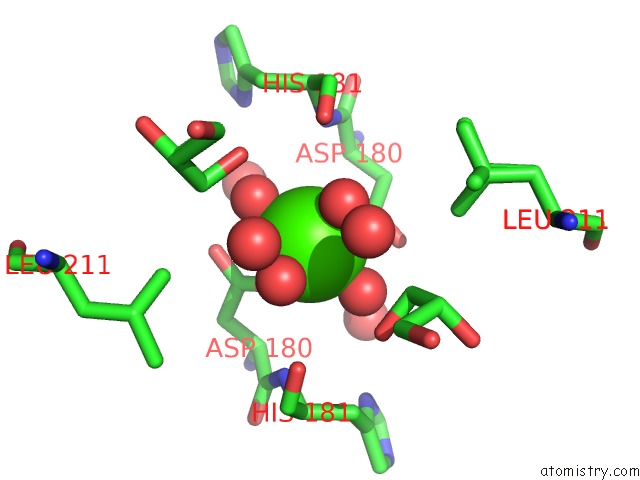
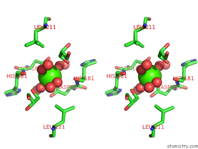
Calcium binding site 1 out of 4 in 4b9b
Go back to
Calcium binding site 1 out
of 4 in the The Structure of the Omega Aminotransferase From Pseudomonas Aeruginosa
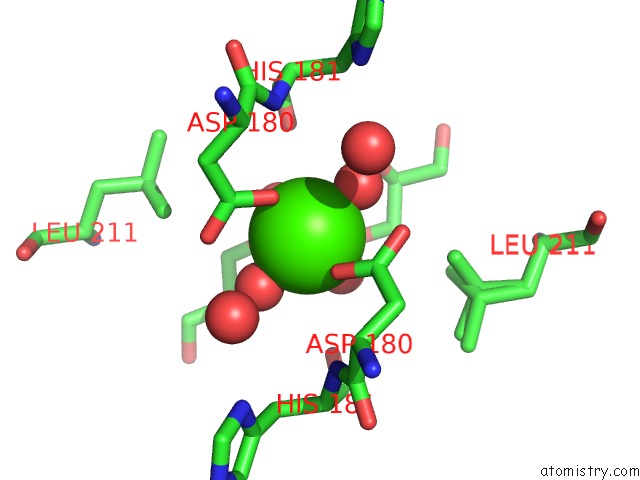
Mono view
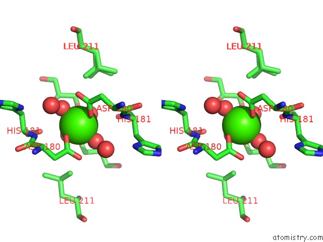
Stereo pair view
Mono view
Stereo pair view
A full contact list of Calcium with other atoms in the Ca binding
site number 1 of The Structure of the Omega Aminotransferase From Pseudomonas Aeruginosa within 5.0Å range:
|
Calcium binding site 2 out of 4 in 4b9b
Go back to
Calcium binding site 2 out
of 4 in the The Structure of the Omega Aminotransferase From Pseudomonas Aeruginosa
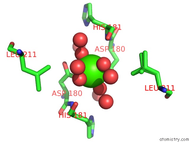
Mono view
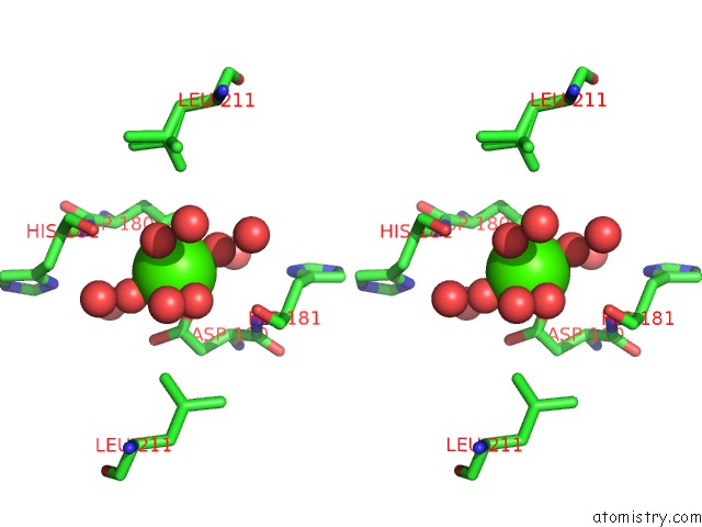
Stereo pair view
Mono view
Stereo pair view
A full contact list of Calcium with other atoms in the Ca binding
site number 2 of The Structure of the Omega Aminotransferase From Pseudomonas Aeruginosa within 5.0Å range:
|
Calcium binding site 3 out of 4 in 4b9b
Go back to
Calcium binding site 3 out
of 4 in the The Structure of the Omega Aminotransferase From Pseudomonas Aeruginosa
Mono view
Stereo pair view
Mono view
Stereo pair view
A full contact list of Calcium with other atoms in the Ca binding
site number 3 of The Structure of the Omega Aminotransferase From Pseudomonas Aeruginosa within 5.0Å range:
|
Calcium binding site 4 out of 4 in 4b9b
Go back to
Calcium binding site 4 out
of 4 in the The Structure of the Omega Aminotransferase From Pseudomonas Aeruginosa

Mono view

Stereo pair view
Mono view
Stereo pair view
A full contact list of Calcium with other atoms in the Ca binding
site number 4 of The Structure of the Omega Aminotransferase From Pseudomonas Aeruginosa within 5.0Å range:
|
Reference:
C.Sayer,
M.N.Isupov,
A.Westlake,
J.A.Littlechild.
Structural Studies with Pseudomonas and Chromobacterium [Omega]-Aminotransferases Provide Insights Into Their Differing Substrate Specificity. Acta Crystallogr.,Sect.D V. 69 564 2013.
ISSN: ISSN 0907-4449
PubMed: 23519665
DOI: 10.1107/S0907444912051670
Page generated: Sat Jul 13 22:38:51 2024
ISSN: ISSN 0907-4449
PubMed: 23519665
DOI: 10.1107/S0907444912051670
Last articles
Zn in 9MJ5Zn in 9HNW
Zn in 9G0L
Zn in 9FNE
Zn in 9DZN
Zn in 9E0I
Zn in 9D32
Zn in 9DAK
Zn in 8ZXC
Zn in 8ZUF