Calcium »
PDB 4bb9-4bu4 »
4bgb »
Calcium in PDB 4bgb: Nucleotide-Bound Closed Form of A Putative Sugar Kinase MK0840 From Methanopyrus Kandleri
Protein crystallography data
The structure of Nucleotide-Bound Closed Form of A Putative Sugar Kinase MK0840 From Methanopyrus Kandleri, PDB code: 4bgb
was solved by
M.Schacherl,
U.Baumann,
with X-Ray Crystallography technique. A brief refinement statistics is given in the table below:
| Resolution Low / High (Å) | 45.147 / 1.34 |
| Space group | P 21 21 21 |
| Cell size a, b, c (Å), α, β, γ (°) | 54.790, 108.540, 117.350, 90.00, 90.00, 90.00 |
| R / Rfree (%) | 15.76 / 18.28 |
Other elements in 4bgb:
The structure of Nucleotide-Bound Closed Form of A Putative Sugar Kinase MK0840 From Methanopyrus Kandleri also contains other interesting chemical elements:
| Magnesium | (Mg) | 4 atoms |
| Potassium | (K) | 5 atoms |
Calcium Binding Sites:
The binding sites of Calcium atom in the Nucleotide-Bound Closed Form of A Putative Sugar Kinase MK0840 From Methanopyrus Kandleri
(pdb code 4bgb). This binding sites where shown within
5.0 Angstroms radius around Calcium atom.
In total only one binding site of Calcium was determined in the Nucleotide-Bound Closed Form of A Putative Sugar Kinase MK0840 From Methanopyrus Kandleri, PDB code: 4bgb:
In total only one binding site of Calcium was determined in the Nucleotide-Bound Closed Form of A Putative Sugar Kinase MK0840 From Methanopyrus Kandleri, PDB code: 4bgb:
Calcium binding site 1 out of 1 in 4bgb
Go back to
Calcium binding site 1 out
of 1 in the Nucleotide-Bound Closed Form of A Putative Sugar Kinase MK0840 From Methanopyrus Kandleri
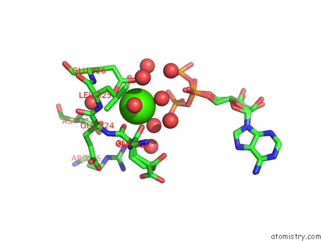
Mono view
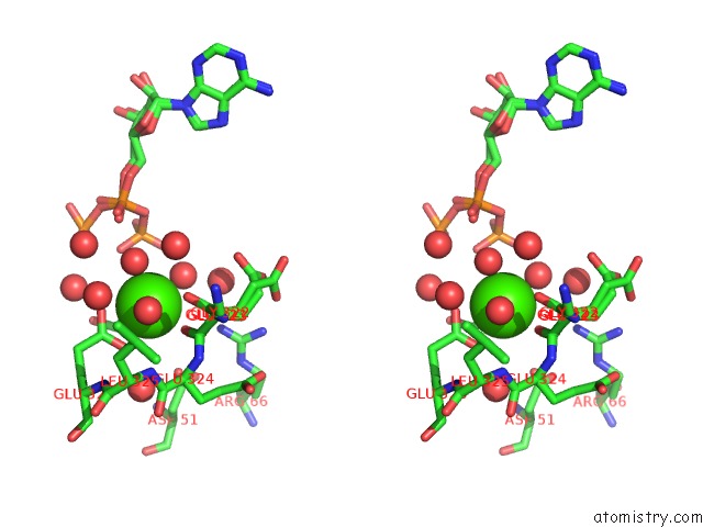
Stereo pair view
Mono view
Stereo pair view
A full contact list of Calcium with other atoms in the Ca binding
site number 1 of Nucleotide-Bound Closed Form of A Putative Sugar Kinase MK0840 From Methanopyrus Kandleri within 5.0Å range:
|
Reference:
M.Schacherl,
S.M.Waltersperger,
U.Baumann.
Structural Characterization of the Ribonuclease H-Like Type Askha Superfamily Kinase MK0840 From Methanopyrus Kandleri Acta Crystallogr.,Sect.D V. 69 2440 2013.
ISSN: ISSN 0907-4449
PubMed: 24311585
DOI: 10.1107/S0907444913022683
Page generated: Sat Jul 13 22:42:18 2024
ISSN: ISSN 0907-4449
PubMed: 24311585
DOI: 10.1107/S0907444913022683
Last articles
Zn in 9MJ5Zn in 9HNW
Zn in 9G0L
Zn in 9FNE
Zn in 9DZN
Zn in 9E0I
Zn in 9D32
Zn in 9DAK
Zn in 8ZXC
Zn in 8ZUF