Calcium »
PDB 4bb9-4bu4 »
4bq2 »
Calcium in PDB 4bq2: Structural Analysis of An Exo-Beta-Agarase
Enzymatic activity of Structural Analysis of An Exo-Beta-Agarase
All present enzymatic activity of Structural Analysis of An Exo-Beta-Agarase:
3.2.1.81;
3.2.1.81;
Protein crystallography data
The structure of Structural Analysis of An Exo-Beta-Agarase, PDB code: 4bq2
was solved by
B.Pluvinage,
J.H.Hehemann,
A.B.Boraston,
with X-Ray Crystallography technique. A brief refinement statistics is given in the table below:
| Resolution Low / High (Å) | 39.28 / 1.90 |
| Space group | P 41 |
| Cell size a, b, c (Å), α, β, γ (°) | 166.666, 166.666, 114.559, 90.00, 90.00, 90.00 |
| R / Rfree (%) | 17.021 / 21.487 |
Calcium Binding Sites:
The binding sites of Calcium atom in the Structural Analysis of An Exo-Beta-Agarase
(pdb code 4bq2). This binding sites where shown within
5.0 Angstroms radius around Calcium atom.
In total 4 binding sites of Calcium where determined in the Structural Analysis of An Exo-Beta-Agarase, PDB code: 4bq2:
Jump to Calcium binding site number: 1; 2; 3; 4;
In total 4 binding sites of Calcium where determined in the Structural Analysis of An Exo-Beta-Agarase, PDB code: 4bq2:
Jump to Calcium binding site number: 1; 2; 3; 4;
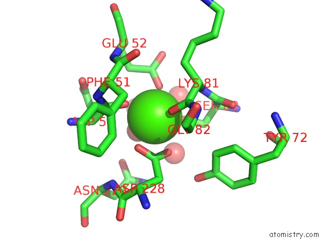
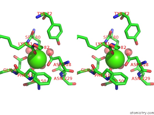
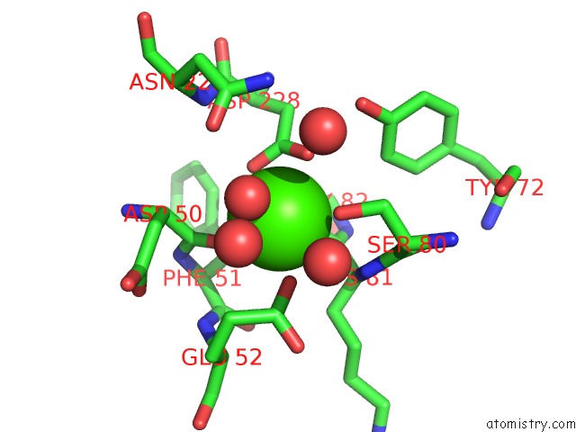
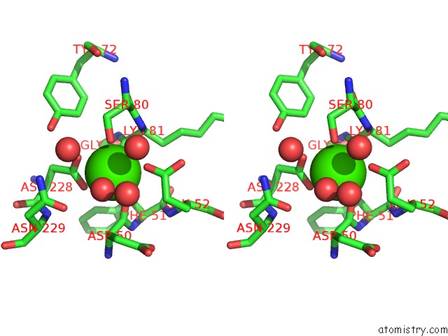
Calcium binding site 1 out of 4 in 4bq2
Go back to
Calcium binding site 1 out
of 4 in the Structural Analysis of An Exo-Beta-Agarase
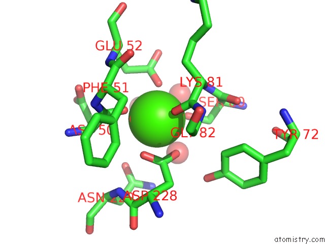
Mono view
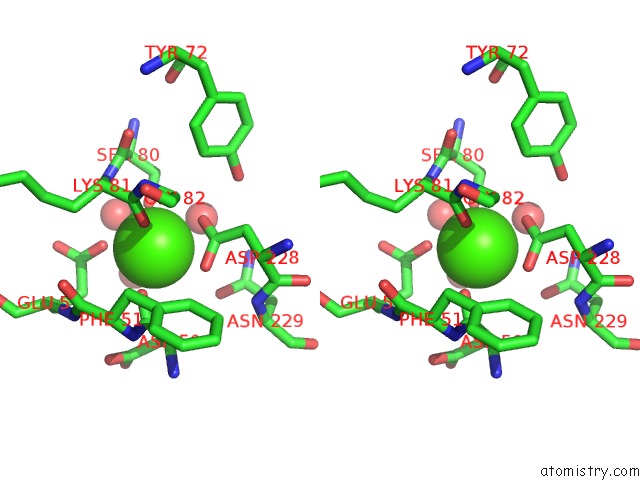
Stereo pair view
Mono view
Stereo pair view
A full contact list of Calcium with other atoms in the Ca binding
site number 1 of Structural Analysis of An Exo-Beta-Agarase within 5.0Å range:
|
Calcium binding site 2 out of 4 in 4bq2
Go back to
Calcium binding site 2 out
of 4 in the Structural Analysis of An Exo-Beta-Agarase
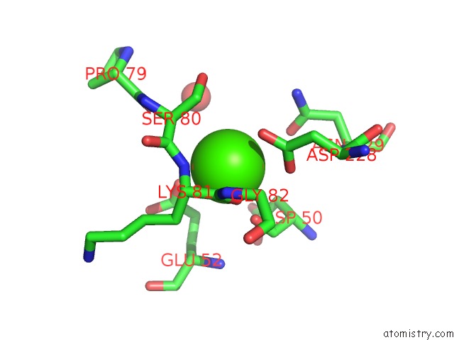
Mono view
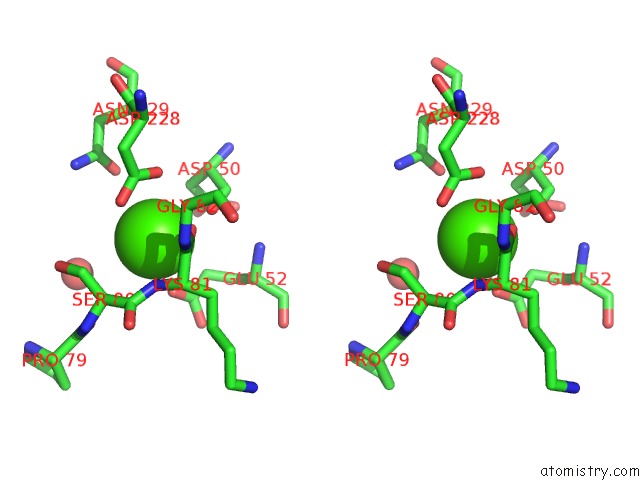
Stereo pair view
Mono view
Stereo pair view
A full contact list of Calcium with other atoms in the Ca binding
site number 2 of Structural Analysis of An Exo-Beta-Agarase within 5.0Å range:
|
Calcium binding site 3 out of 4 in 4bq2
Go back to
Calcium binding site 3 out
of 4 in the Structural Analysis of An Exo-Beta-Agarase
Mono view
Stereo pair view
Mono view
Stereo pair view
A full contact list of Calcium with other atoms in the Ca binding
site number 3 of Structural Analysis of An Exo-Beta-Agarase within 5.0Å range:
|
Calcium binding site 4 out of 4 in 4bq2
Go back to
Calcium binding site 4 out
of 4 in the Structural Analysis of An Exo-Beta-Agarase
Mono view
Stereo pair view
Mono view
Stereo pair view
A full contact list of Calcium with other atoms in the Ca binding
site number 4 of Structural Analysis of An Exo-Beta-Agarase within 5.0Å range:
|
Reference:
B.Pluvinage,
J.H.Hehemann,
A.B.Boraston.
Substrate Recognition and Hydrolysis By A Family 50 Exo-Beta-Agarase AGA50D From the Marine Bacterium Saccharophagus Degradans J.Biol.Chem. V. 288 28078 2013.
ISSN: ISSN 0021-9258
PubMed: 23921382
DOI: 10.1074/JBC.M113.491068
Page generated: Sat Jul 13 22:47:11 2024
ISSN: ISSN 0021-9258
PubMed: 23921382
DOI: 10.1074/JBC.M113.491068
Last articles
Zn in 9J0NZn in 9J0O
Zn in 9J0P
Zn in 9FJX
Zn in 9EKB
Zn in 9C0F
Zn in 9CAH
Zn in 9CH0
Zn in 9CH3
Zn in 9CH1