Calcium »
PDB 4bw8-4cg0 »
4c0k »
Calcium in PDB 4c0k: Crystal Structure of Drosophila Miro Ef Hand and Cgtpase Domains Bound to One Calcium Ion (Ca-Miros)
Protein crystallography data
The structure of Crystal Structure of Drosophila Miro Ef Hand and Cgtpase Domains Bound to One Calcium Ion (Ca-Miros), PDB code: 4c0k
was solved by
J.L.Klosowiak,
P.J.Focia,
Z.Wawrzak,
S.Chakravarthy,
E.C.Landahl,
D.M.Freymann,
S.E.Rice,
with X-Ray Crystallography technique. A brief refinement statistics is given in the table below:
| Resolution Low / High (Å) | 42.075 / 2.80 |
| Space group | P 32 2 1 |
| Cell size a, b, c (Å), α, β, γ (°) | 82.240, 82.240, 156.441, 90.00, 90.00, 120.00 |
| R / Rfree (%) | 20.72 / 24.22 |
Other elements in 4c0k:
The structure of Crystal Structure of Drosophila Miro Ef Hand and Cgtpase Domains Bound to One Calcium Ion (Ca-Miros) also contains other interesting chemical elements:
| Sodium | (Na) | 1 atom |
Calcium Binding Sites:
The binding sites of Calcium atom in the Crystal Structure of Drosophila Miro Ef Hand and Cgtpase Domains Bound to One Calcium Ion (Ca-Miros)
(pdb code 4c0k). This binding sites where shown within
5.0 Angstroms radius around Calcium atom.
In total only one binding site of Calcium was determined in the Crystal Structure of Drosophila Miro Ef Hand and Cgtpase Domains Bound to One Calcium Ion (Ca-Miros), PDB code: 4c0k:
In total only one binding site of Calcium was determined in the Crystal Structure of Drosophila Miro Ef Hand and Cgtpase Domains Bound to One Calcium Ion (Ca-Miros), PDB code: 4c0k:
Calcium binding site 1 out of 1 in 4c0k
Go back to
Calcium binding site 1 out
of 1 in the Crystal Structure of Drosophila Miro Ef Hand and Cgtpase Domains Bound to One Calcium Ion (Ca-Miros)
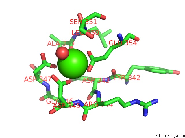
Mono view
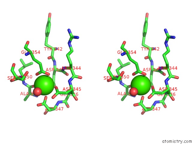
Stereo pair view
Mono view
Stereo pair view
A full contact list of Calcium with other atoms in the Ca binding
site number 1 of Crystal Structure of Drosophila Miro Ef Hand and Cgtpase Domains Bound to One Calcium Ion (Ca-Miros) within 5.0Å range:
|
Reference:
J.L.Klosowiak,
P.J.Focia,
S.Chakravarthy,
E.C.Landahl,
D.M.Freymann,
S.E.Rice.
Structural Coupling of the Ef Hand and C-Terminal Gtpase Domains in the Mitochondrial Protein Miro. Embo Rep. V. 14 968 2013.
ISSN: ISSN 1469-221X
PubMed: 24071720
DOI: 10.1038/EMBOR.2013.151
Page generated: Tue Jul 8 19:07:08 2025
ISSN: ISSN 1469-221X
PubMed: 24071720
DOI: 10.1038/EMBOR.2013.151
Last articles
Cl in 5ICWCl in 5IDY
Cl in 5IEQ
Cl in 5IDN
Cl in 5ICR
Cl in 5IBP
Cl in 5ICU
Cl in 5ICP
Cl in 5IA3
Cl in 5IAE