Calcium »
PDB 4bw8-4cg0 »
4caj »
Calcium in PDB 4caj: Crystallographic Structure of the Mouse Sign-R1 Crd Domain in Complex with Sialic Acid
Protein crystallography data
The structure of Crystallographic Structure of the Mouse Sign-R1 Crd Domain in Complex with Sialic Acid, PDB code: 4caj
was solved by
N.Silva-Martin,
S.G.Bartual,
J.A.Hermoso,
with X-Ray Crystallography technique. A brief refinement statistics is given in the table below:
| Resolution Low / High (Å) | 14.948 / 2.19 |
| Space group | C 1 2 1 |
| Cell size a, b, c (Å), α, β, γ (°) | 146.200, 94.470, 76.140, 90.00, 121.42, 90.00 |
| R / Rfree (%) | 20.53 / 28.12 |
Other elements in 4caj:
The structure of Crystallographic Structure of the Mouse Sign-R1 Crd Domain in Complex with Sialic Acid also contains other interesting chemical elements:
| Chlorine | (Cl) | 2 atoms |
Calcium Binding Sites:
The binding sites of Calcium atom in the Crystallographic Structure of the Mouse Sign-R1 Crd Domain in Complex with Sialic Acid
(pdb code 4caj). This binding sites where shown within
5.0 Angstroms radius around Calcium atom.
In total 4 binding sites of Calcium where determined in the Crystallographic Structure of the Mouse Sign-R1 Crd Domain in Complex with Sialic Acid, PDB code: 4caj:
Jump to Calcium binding site number: 1; 2; 3; 4;
In total 4 binding sites of Calcium where determined in the Crystallographic Structure of the Mouse Sign-R1 Crd Domain in Complex with Sialic Acid, PDB code: 4caj:
Jump to Calcium binding site number: 1; 2; 3; 4;
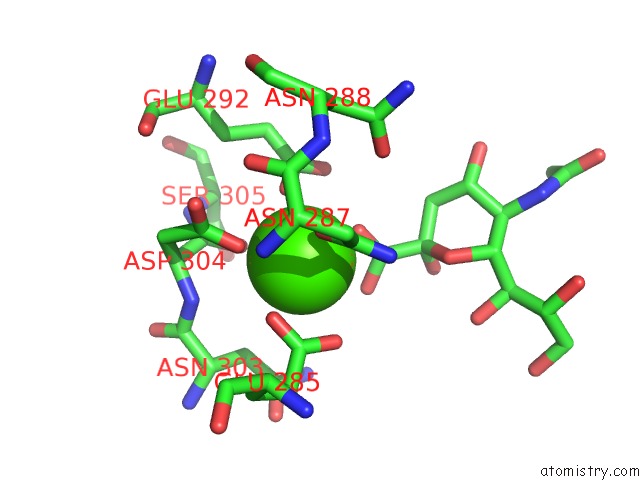
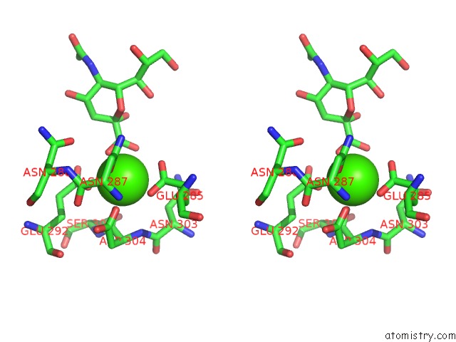
Calcium binding site 1 out of 4 in 4caj
Go back to
Calcium binding site 1 out
of 4 in the Crystallographic Structure of the Mouse Sign-R1 Crd Domain in Complex with Sialic Acid
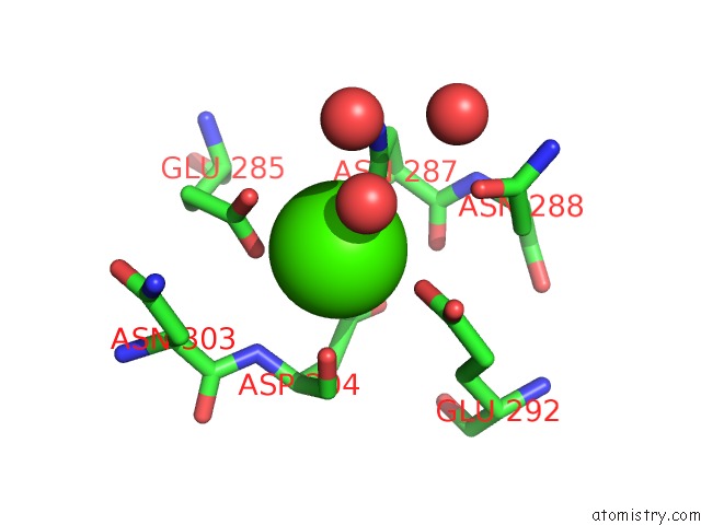
Mono view
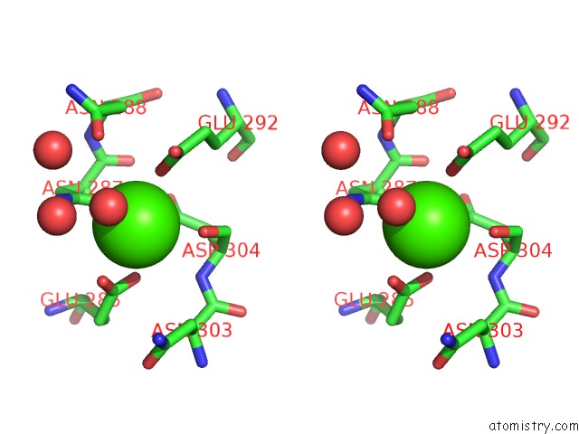
Stereo pair view
Mono view
Stereo pair view
A full contact list of Calcium with other atoms in the Ca binding
site number 1 of Crystallographic Structure of the Mouse Sign-R1 Crd Domain in Complex with Sialic Acid within 5.0Å range:
|
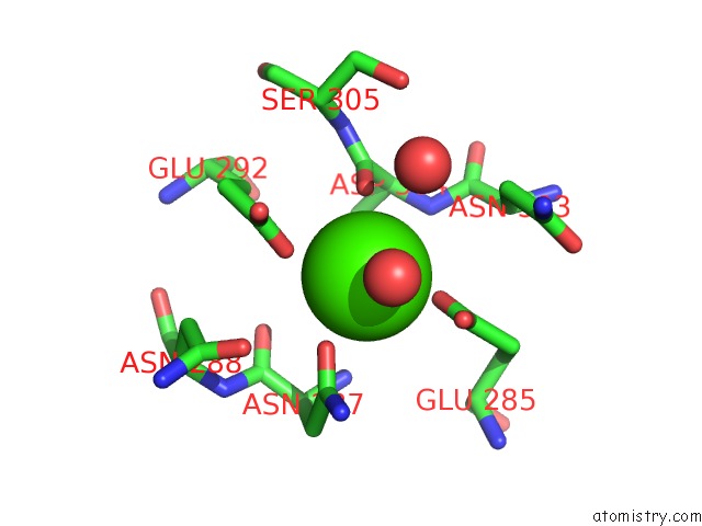
Calcium binding site 2 out of 4 in 4caj
Go back to
Calcium binding site 2 out
of 4 in the Crystallographic Structure of the Mouse Sign-R1 Crd Domain in Complex with Sialic Acid
Mono view
Stereo pair view
Mono view
Stereo pair view
A full contact list of Calcium with other atoms in the Ca binding
site number 2 of Crystallographic Structure of the Mouse Sign-R1 Crd Domain in Complex with Sialic Acid within 5.0Å range:
|
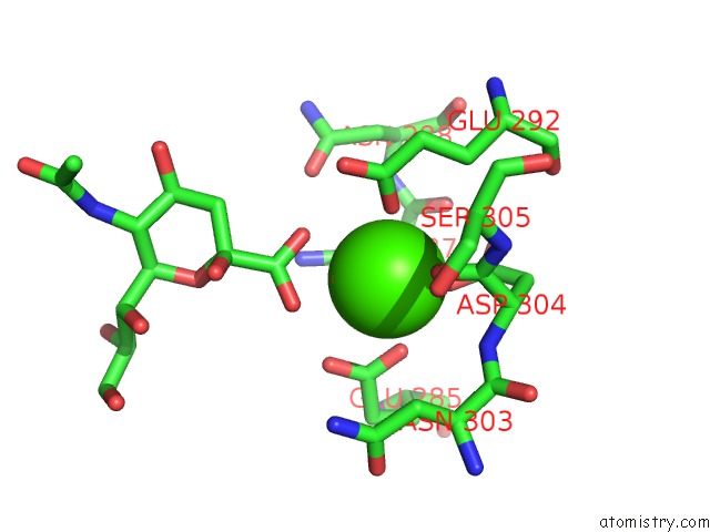
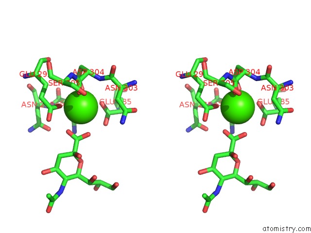
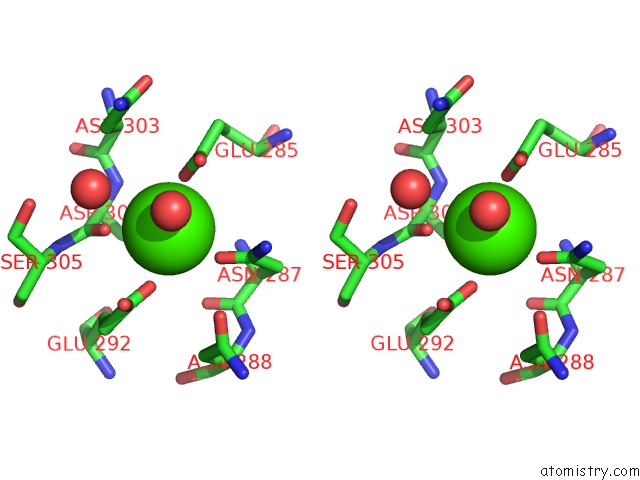
Calcium binding site 3 out of 4 in 4caj
Go back to
Calcium binding site 3 out
of 4 in the Crystallographic Structure of the Mouse Sign-R1 Crd Domain in Complex with Sialic Acid
Mono view
Stereo pair view
Mono view
Stereo pair view
A full contact list of Calcium with other atoms in the Ca binding
site number 3 of Crystallographic Structure of the Mouse Sign-R1 Crd Domain in Complex with Sialic Acid within 5.0Å range:
|
Calcium binding site 4 out of 4 in 4caj
Go back to
Calcium binding site 4 out
of 4 in the Crystallographic Structure of the Mouse Sign-R1 Crd Domain in Complex with Sialic Acid
Mono view
Stereo pair view
Mono view
Stereo pair view
A full contact list of Calcium with other atoms in the Ca binding
site number 4 of Crystallographic Structure of the Mouse Sign-R1 Crd Domain in Complex with Sialic Acid within 5.0Å range:
|
Reference:
N.Silva-Martin,
S.G.Bartual,
A.Rodriguez,
E.Ramirez,
P.Chacon,
R.M.Anthony,
C.G.Park,
J.A.Hermoso.
Structural Basis For Selective Recognition of Endogenous and Microbial Polysaccharides By Macrophage Receptor Sign-R1 Structure 2014.
ISSN: ESSN 1878-4186
DOI: 10.1016/J.STR.2014.09.001
Page generated: Tue Jul 8 19:08:42 2025
ISSN: ESSN 1878-4186
DOI: 10.1016/J.STR.2014.09.001
Last articles
Cl in 5L64Cl in 5L63
Cl in 5L5Z
Cl in 5L61
Cl in 5L5Y
Cl in 5L60
Cl in 5L5W
Cl in 5L5X
Cl in 5L5V
Cl in 5L5U