Calcium »
PDB 4bw8-4cg0 »
4cbw »
Calcium in PDB 4cbw: Crystal Structure of Plasmodium Berghei Actin I with D-Loop From Muscle Actin
Protein crystallography data
The structure of Crystal Structure of Plasmodium Berghei Actin I with D-Loop From Muscle Actin, PDB code: 4cbw
was solved by
J.Vahokoski,
S.P.Bhargav,
A.Desfosses,
M.Andreadaki,
E.P.Kumpula,
A.Ignatev,
S.Munico Martinez,
S.Lepper,
F.Frischknecht,
I.Siden-Kiamos,
C.Sachse,
I.Kursula,
with X-Ray Crystallography technique. A brief refinement statistics is given in the table below:
| Resolution Low / High (Å) | 38.581 / 2.50 |
| Space group | P 21 21 21 |
| Cell size a, b, c (Å), α, β, γ (°) | 54.240, 69.530, 178.830, 90.00, 90.00, 90.00 |
| R / Rfree (%) | 21.07 / 26.4 |
Calcium Binding Sites:
The binding sites of Calcium atom in the Crystal Structure of Plasmodium Berghei Actin I with D-Loop From Muscle Actin
(pdb code 4cbw). This binding sites where shown within
5.0 Angstroms radius around Calcium atom.
In total 3 binding sites of Calcium where determined in the Crystal Structure of Plasmodium Berghei Actin I with D-Loop From Muscle Actin, PDB code: 4cbw:
Jump to Calcium binding site number: 1; 2; 3;
In total 3 binding sites of Calcium where determined in the Crystal Structure of Plasmodium Berghei Actin I with D-Loop From Muscle Actin, PDB code: 4cbw:
Jump to Calcium binding site number: 1; 2; 3;
Calcium binding site 1 out of 3 in 4cbw
Go back to
Calcium binding site 1 out
of 3 in the Crystal Structure of Plasmodium Berghei Actin I with D-Loop From Muscle Actin

Mono view
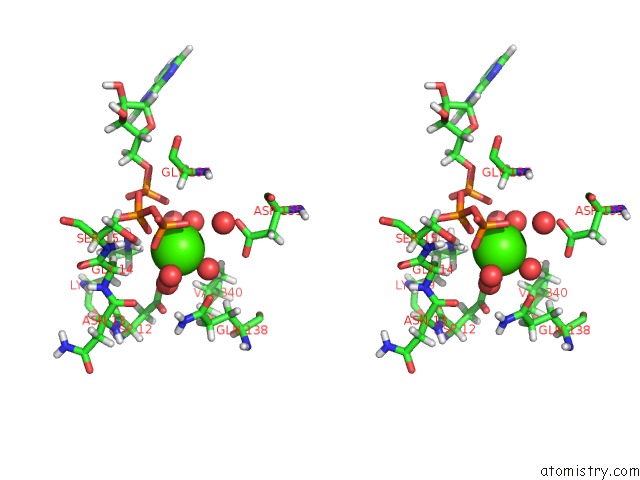
Stereo pair view
Mono view
Stereo pair view
A full contact list of Calcium with other atoms in the Ca binding
site number 1 of Crystal Structure of Plasmodium Berghei Actin I with D-Loop From Muscle Actin within 5.0Å range:
|
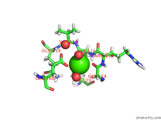
Calcium binding site 2 out of 3 in 4cbw
Go back to
Calcium binding site 2 out
of 3 in the Crystal Structure of Plasmodium Berghei Actin I with D-Loop From Muscle Actin
Mono view

Stereo pair view
Mono view
Stereo pair view
A full contact list of Calcium with other atoms in the Ca binding
site number 2 of Crystal Structure of Plasmodium Berghei Actin I with D-Loop From Muscle Actin within 5.0Å range:
|
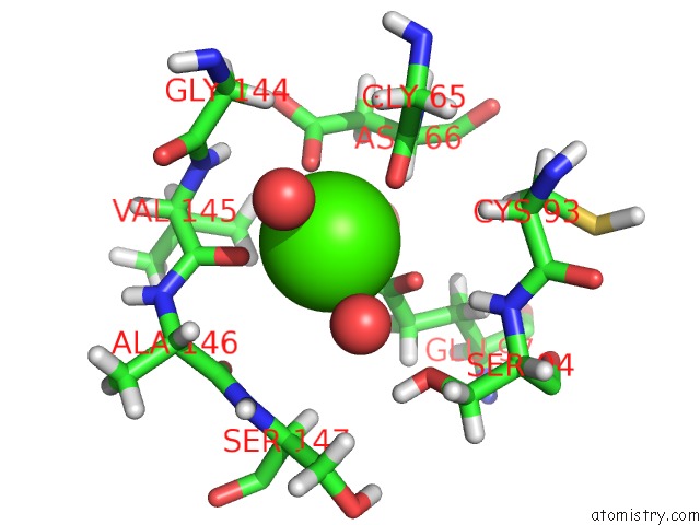
Calcium binding site 3 out of 3 in 4cbw
Go back to
Calcium binding site 3 out
of 3 in the Crystal Structure of Plasmodium Berghei Actin I with D-Loop From Muscle Actin
Mono view

Stereo pair view
Mono view
Stereo pair view
A full contact list of Calcium with other atoms in the Ca binding
site number 3 of Crystal Structure of Plasmodium Berghei Actin I with D-Loop From Muscle Actin within 5.0Å range:
|
Reference:
J.Vahokoski,
S.P.Bhargav,
A.Desfosses,
M.Andreadaki,
E.Kumpula,
S.M.Martinez,
A.Ignatev,
S.Lepper,
F.Frischknecht,
I.Siden-Kiamos,
C.Sachse,
I.Kursula.
Structural Differences Explain Diverse Functions of Plasmodium Actins. Plos Pathog. V. 10 4091 2014.
ISSN: ISSN 1553-7366
PubMed: 24743229
DOI: 10.1371/JOURNAL.PPAT.1004091
Page generated: Sat Jul 13 23:01:10 2024
ISSN: ISSN 1553-7366
PubMed: 24743229
DOI: 10.1371/JOURNAL.PPAT.1004091
Last articles
Zn in 9J0NZn in 9J0O
Zn in 9J0P
Zn in 9FJX
Zn in 9EKB
Zn in 9C0F
Zn in 9CAH
Zn in 9CH0
Zn in 9CH3
Zn in 9CH1